Key Takeaways
- Elastic Cartilage provides flexible support in regions requiring elasticity, unlike Hyaline Cartilage which offers firm, smooth surfaces.
- Hyaline Cartilage primarily composes the embryonic skeleton and covers joint surfaces, whereas Elastic Cartilage are found in structures needing resilience and flexibility.
- The composition of Elastic Cartilage includes elastic fibers that give it greater bendability, while Hyaline Cartilage is mainly made of a dense collagen matrix.
- Healing and regeneration rates differ; Elastic Cartilage tends to heal slower due to its complex fiber network compared to Hyaline Cartilage’s relatively quicker repair process.
- Both types of cartilage play vital roles in their respective locations, but their structural differences suit their functions in the body’s architecture.
What is Elastic Cartilage?
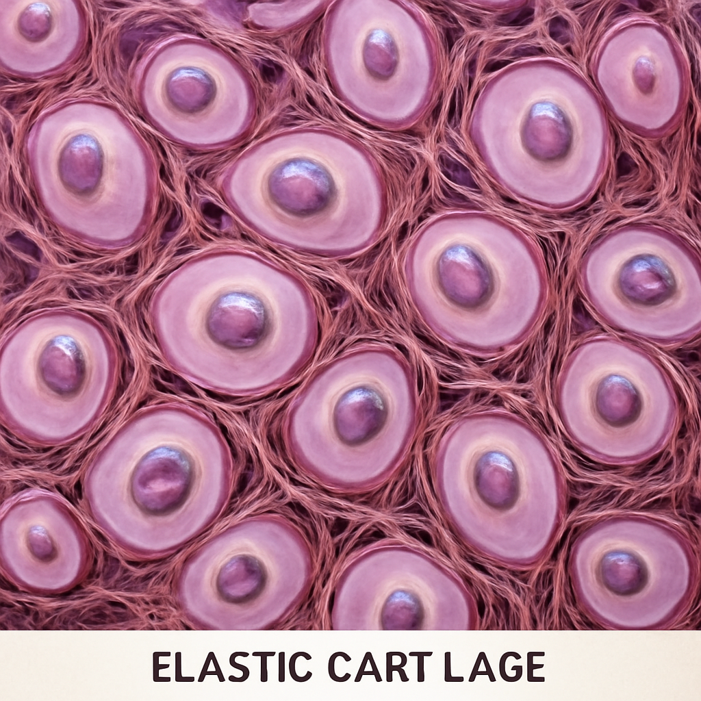
Elastic Cartilage is a specialized form of cartilage characterized by its high content of elastic fibers, allowing it to bend and return to shape. It is found in locations where flexibility and resilience are needed in the body’s framework.
Structural Composition and Fiber Network
Elastic Cartilage contains a dense network of elastic fibers embedded within a gel-like ground substance. This fiber network grants it elasticity, enabling structures to stretch and recoil without damage. The elastic fibers are interwoven with collagen fibers, providing both flexibility and durability.
This composition distinguishes it from other cartilage types, primarily because of the prominence of elastic fibers. These fibers are made of elastin, a protein that imparts the stretching ability. The ground substance surrounding these fibers maintains the tissue’s resilience, keeping it supple yet supportive,
In the context of biomechanical function, this fiber arrangement allows structures to withstand frequent bending and deformation. For example, in the ear, elastic fibers help maintain shape while accommodating movement, which is crucial for auditory functions.
The elasticity also plays a role in the respiratory system, particularly in the epiglottis, where it allows the structure to move and fold during swallowing. Although incomplete. This flexibility is vital in preventing food from entering the windpipe and ensuring smooth airway function.
Locations and Functional Significance
Elastic Cartilage is prominently located in the external ear, forming the flexible framework of the auricle. Its ability to bend and retain shape makes it ideal for this purpose, providing both support and pliability.
Another key site is the epiglottis, which acts as a lid over the larynx during swallowing. Its elastic nature ensures that it can move swiftly and return to its original position after movement, protecting the airway.
The auditory tube, connecting the middle ear to the pharynx, also contains elastic cartilage. This allows the tube to open and close as necessary, aiding pressure regulation and sound transmission.
In the larynx, elastic cartilage forms the cuneiform and corniculate cartilages, which support the vocal cords and assist in voice modulation. Although incomplete. Its flexibility is crucial for speech and breathing functions.
By contrast, elastic cartilage’s resilience is vital in regions that experience frequent movement, preventing permanent deformation and ensuring structural integrity over time.
Development and Regeneration
Elastic Cartilage develops from mesenchymal stem cells during fetal growth, closely associated with cartilage precursor cells. Its formation involves a process called chondrogenesis, where these cells produce the characteristic elastic fibers and cartilage matrix.
The regeneration of elastic cartilage is slower compared to other tissue types, partly because of its dense elastic fiber network. Damage in areas like the ear often requires surgical intervention for repair, as natural healing can be limited.
In cases of injury, the elastic fibers’ complex arrangement complicates the healing process, sometimes leading to scar tissue formation instead of true cartilage regeneration. This factor influences clinical approaches to injuries involving elastic cartilage.
Research explores tissue engineering techniques, such as scaffolding and stem cell therapy, to enhance elastic cartilage repair. These methods aim to mimic the natural elasticity and support the regeneration process more effectively,
Understanding the development pathways and regenerative limitations of elastic cartilage helps in devising better treatment strategies for injuries and congenital deformities involving elastic structures.
Clinical Relevance and Disorders
Elastic Cartilage can be affected by trauma, infection, or congenital deformities, often resulting in deformities of the ear or airway obstructions. Conditions like auricular hematoma or cartilage necrosis are examples of clinical issues involving elastic cartilage.
In reconstructive surgeries, grafts using elastic cartilage are common for restoring ear shape, especially in cases of traumatic loss or congenital anomalies like microtia. Although incomplete. Surgeons carefully preserve the elastic fiber network to maintain functionality.
In the respiratory pathway, damage to elastic cartilage in the epiglottis can lead to swallowing difficulties or airway obstruction, necessitating surgical correction or tissue grafts.
Allergic reactions or autoimmune conditions rarely target elastic cartilage specifically, but when they do, they can cause inflammation and deformity, impacting breathing or speech.
Advancements in biomaterials and regenerative medicine continue to improve outcomes for disorders involving elastic cartilage, emphasizing minimally invasive repair and tissue compatibility.
What is Hyaline Cartilage?
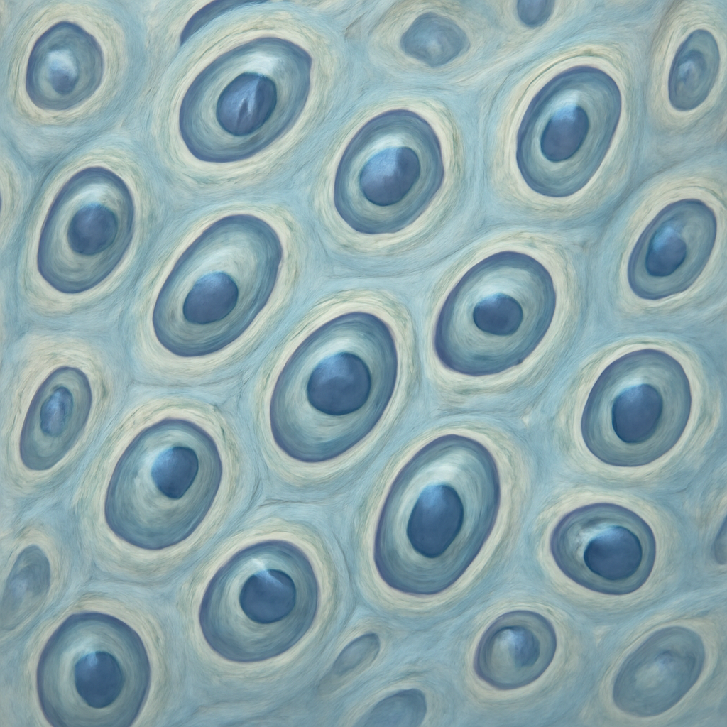
Hyaline Cartilage is a smooth, glassy connective tissue that covers joint surfaces and provides structural support during development. It is the most abundant cartilage type found in the human body,
Microstructure and Composition
Hyaline Cartilage contains a dense matrix rich in type II collagen fibers, which give it strength and flexibility without sacrificing smoothness. The collagen fibers is embedded in a gel-like ground substance composed of proteoglycans and water, providing resilience.
The collagen network is arranged in a way that maintains a smooth surface, essential for low-friction articulation. The high water content allows it to absorb shock and distribute loads evenly across joint surfaces.
Unlike elastic cartilage, hyaline cartilage has few elastic fibers; instead, it relies on its collagen framework for structural integrity. This makes it less flexible but more durable under compressive forces.
The chondrocytes, or cartilage cells, are embedded within small spaces called lacunae in the matrix. They produce and maintain the extracellular components, ensuring the tissue’s function over time.
Distribution and Functional Roles
Hyaline cartilage covers articular surfaces of bones in synovial joints, facilitating smooth movement and reducing friction. It is also found in the respiratory tract, forming the cartilage rings of the trachea and bronchi.
During fetal development, hyaline cartilage serves as a precursor for the formation of most bones through endochondral ossification. It provides a temporary scaffold that is replaced by bone as growth proceeds.
In the nasal septum, hyaline cartilage maintains the shape and support of the nose structure. Its resilience allows it to withstand constant exposure to air and mechanical stresses.
In the larynx, hyaline cartilage forms the thyroid and cricoid cartilages, which support voice production and airway maintenance. Its durability is vital for these protective functions.
In joints, hyaline cartilage’s smooth surface and shock absorption capabilities are essential for mobility and load bearing, making it central to skeletal movement.
Growth and Repair Mechanisms
Hyaline cartilage grows by both appositional and interstitial mechanisms, with chondroblasts adding new layers at the surface and chondrocytes dividing within the matrix. This dual growth method helps in shaping the skeletal elements.
Its repair capacity is limited because cartilage lacks blood vessels, relying on diffusion for nutrient supply. Damage often results in slow healing or formation of fibrocartilage instead of true hyaline cartilage.
In degenerative conditions like osteoarthritis, hyaline cartilage deteriorates, leading to joint pain and reduced mobility. The inability to regenerate effectively challenges clinical management.
Research into stimulating chondrocyte activity or introducing tissue-engineered cartilage aims to improve repair processes, especially for joint injuries.
The avascular nature of hyaline cartilage is a double-edged sword: it preserves the tissue but complicates healing, necessitating innovative solutions for restoration.
Pathological Conditions and Treatments
Degeneration of hyaline cartilage causes osteoarthritis, characterized by joint pain, stiffness, and loss of function. The condition arises from wear and tear, inflammation, or injury.
Injury to hyaline cartilage often results in persistent pain because of its low regenerative capacity, requiring surgical procedures like microfracture or cartilage grafting for repair.
Autoimmune diseases like rheumatoid arthritis can target hyaline cartilage, leading to erosion and joint deformity. Managing inflammation is critical to prevent further damage.
In some cases, hyaline cartilage damage is managed with pharmacological agents, physical therapy, or joint replacements, depending on severity. Regenerative medicine approaches are increasingly being explored for long-term solutions.
Understanding the biochemical pathways involved in hyaline cartilage degradation is key to developing targeted therapies for joint preservation and restoration.
Comparison Table
Below is a detailed comparison between Elastic Cartilage and Hyaline Cartilage based on several aspects.
| Parameter of Comparison | Elastic Cartilage | Hyaline Cartilage | |
|---|---|---|---|
| Fiber Composition | Rich in elastic fibers that enable flexibility | Primarily collagen type II fibers, providing strength and smoothness | |
| Location | External ear, epiglottis, larynx, auditory tubes | Articular surfaces, nasal septum, trachea, fetal skeleton | |
| Mechanical Properties | Highly flexible, elastic, capable of bending without damage | Resists compression, provides smooth articulation | |
| Healing Capacity | Slower regeneration, often needs surgical intervention | Limited healing due to avascularity, slow repair | |
| Support Function | Supports structures requiring flexibility, like the ear and larynx | Provides structural support and smooth surfaces in joints and respiratory tract | |
| Matrix Composition | Elastic fibers embedded in a gel-like ground substance | Dense collagen with proteoglycans and water | |
| Elasticity | High elasticity, enables bending and recoil | Minimal elasticity, mainly resists compressive forces | |
| Developmental Role | Supports structures that change shape, like the ear | Precursor to bone in fetal development, supports joint surfaces | |
| Common Injuries | Deformities from trauma, damage affecting the ear or airway | Cartilage erosion in osteoarthritis, joint injuries | |
| Clinical Uses | Grafts in reconstructive ear surgeries, airway repairs | Cartilage repair in joints, nasal reconstructions |
Key Differences
Here are the most distinct features separating Elastic Cartilage from Hyaline Cartilage:
- Fiber Content — Elastic Cartilage contains elastic fibers that allow bending, whereas Hyaline Cartilage is mainly composed of collagen type II fibers that provide rigidity and smoothness.
- Structural Location — Elastic Cartilage supports flexible structures like the ear and epiglottis, while Hyaline Cartilage is found on joint surfaces and in developing bones.
- Flexibility versus Strength — Elastic Cartilage is highly bendable, while Hyaline Cartilage offers resistance to compression and wear.
- Healing Rate — Elastic Cartilage heals slower due to complex fiber networks, in contrast to Hyaline Cartilage, which has limited but somewhat more efficient repair mechanisms.
- Matrix Composition — The ground substance in Elastic Cartilage is rich in elastic fibers, while Hyaline Cartilage’s matrix is dominated by collagen fibers and proteoglycans.
- Functional Role — Elastic Cartilage maintains shape and flexibility of structures, whereas Hyaline Cartilage facilitates smooth joint movements and supports development.
- Injury Response — Elastic Cartilage is more susceptible to deformation and requires surgical intervention, while Hyaline Cartilage degeneration leads to joint diseases like osteoarthritis.
FAQs
Can Elastic Cartilage regenerate after severe injury?
Regeneration of Elastic Cartilage is limited because of its dense elastic fiber network and avascular nature. When severely damaged, it often requires surgical procedures for repair, and spontaneous regeneration is rare.
Is Hyaline Cartilage capable of healing itself after damage?
Hyaline Cartilage has minimal regenerative capacity due to its lack of blood vessels, making natural healing slow or incomplete. This is why injuries often lead to persistent joint issues and sometimes require medical intervention.
What makes Elastic Cartilage more flexible than other cartilage types?
The abundance of elastic fibers within Elastic Cartilage allows it to bend and recoil without losing shape, providing resilience in dynamic structures like the ear and larynx, unlike the more rigid Hyaline Cartilage.
How do the functions of these cartilages impact clinical treatments?
Elastic Cartilage’s flexibility makes it suitable for reconstructive grafts in the ear and airway, while Hyaline Cartilage’s durability is critical in joint repair and prosthetic applications, guiding different surgical approaches.