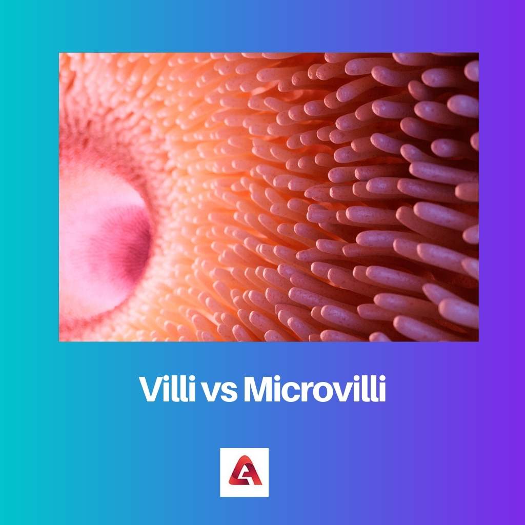
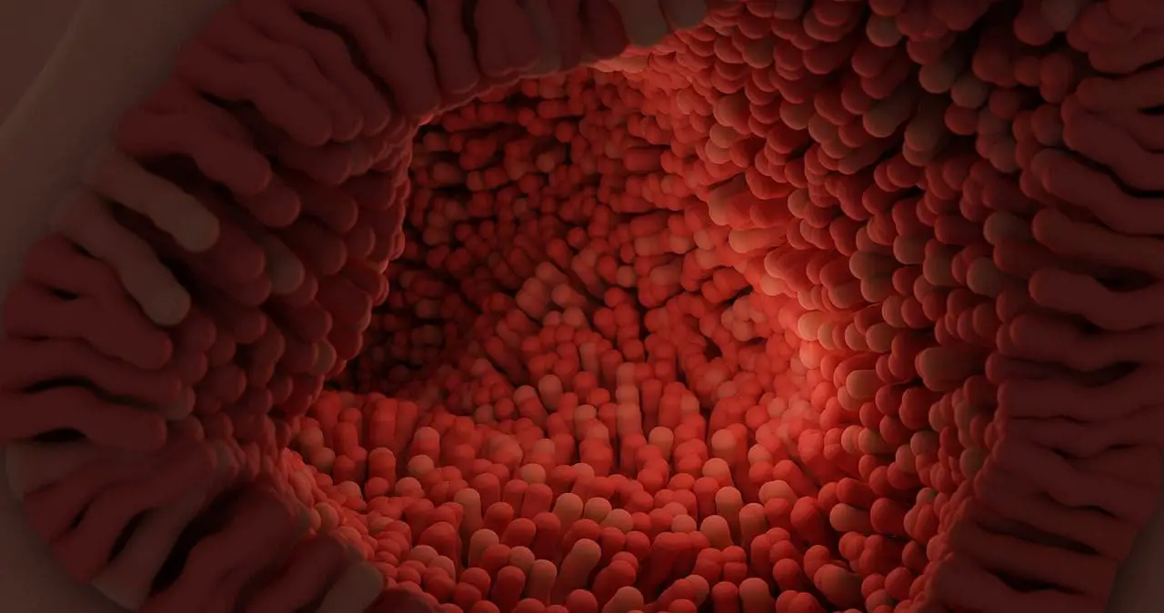
Villi are finger-like projections found in the small intestine that increase its surface area for nutrient absorption. Microvilli are even smaller protrusions on the surface of villi cells, further enhancing absorption by increasing surface area and containing enzymes for final digestion.
Key Takeaways
- Villi are finger-like projections found in the small intestine, while microvilli are even smaller projections on the surface of each villus.
- Villi increase the surface area of the small intestine for nutrient absorption, while microvilli increase the surface area even further.
- Villi are visible to the naked eye, while microvilli can only be seen under a microscope.
Villi vs Microvilli
Vali is finger-like projections in the lining of the small intestine that increase the intestine’s surface area for efficient absorption of nutrients. Microvilli are small finger-like projections found on the surface of certain cells, made up of bundles of actin filaments.

Villi are big finger-like projections in the walls of the small intestines that extend to the lumen. They have a length of 0.5 to 1.6 millimetres. Their main function is to increase the surface area to absorb the various nutrients that reach the small intestines.
Microvilli are cellular structures that are not only found on top of villi but also on many other organs to help them with their functions. They are found only on the cell membrane of epithermal cells. Microvilli are minute projections, much like the villi, but smaller.
Comparison Table
| Feature | Villi | Microvilli |
|---|---|---|
| Location | Inner lining of small intestine | Surface of epithelial cells in various organs |
| Size | Finger-like projections, visible to naked eye | Tiny brush-like projections, microscopic |
| Structure | Composed of epithelial cells, capillaries, and lacteals | Composed of actin filaments and a plasma membrane |
| Function | Increase surface area for nutrient absorption | Increase surface area for absorption and secretion |
| Examples | Abundant in the small intestine | Found in the small intestine, kidney tubules, respiratory tract |
What is Villi?
Villi are microscopic, finger-like projections that line the inner surface of the small intestine, playing a crucial role in the absorption of nutrients from digested food. Structurally, they resemble tiny hair-like structures, densely covering the intestinal lining. Their presence significantly increases the surface area available for nutrient absorption, facilitating efficient nutrient uptake by the body.
Structure of Villi
Villi consist of a central core of connective tissue, surrounded by a layer of epithelial cells. This core contains blood vessels, lymphatic vessels (known as lacteals), and nerves. The outer layer of epithelial cells is specialized for absorption, featuring microvilli on their surface, which further amplify the absorptive capacity of the villi.
Function of Villi
- Increased Surface Area: The primary function of villi is to increase the surface area of the small intestine for nutrient absorption. This increased surface area allows for more efficient absorption of nutrients, including carbohydrates, proteins, fats, vitamins, and minerals, into the bloodstream.
- Nutrient Absorption: Villi are equipped with specialized cells, such as enterocytes, which are responsible for absorbing nutrients from the digested food passing through the small intestine. These cells transport absorbed nutrients across their membranes into the bloodstream or lymphatic system.
- Secretion of Enzymes and Mucus: Apart from absorption, villi also play a role in the secretion of enzymes and mucus. Enzymes released by the cells of the villi aid in the final stages of digestion, breaking down complex nutrients into simpler molecules for absorption. Mucus secreted by goblet cells helps lubricate the intestinal lining and protect it from mechanical damage.
- Immune Function: Villi contribute to the immune function of the intestine by harboring immune cells such as lymphocytes, which help defend against harmful pathogens that may enter the digestive tract.

What is Microvilli?
Microvilli are tiny, hair-like protrusions found on the surface of epithelial cells that line the small intestine and other areas of the body. They are essential for increasing the surface area available for absorption and play a crucial role in nutrient uptake and cellular function.
Structure of Microvilli
Microvilli are structurally composed of actin filaments, which are part of the cell’s cytoskeleton, extending into the core of each microvillus. These actin filaments give microvilli their characteristic shape and provide structural support. Each microvillus is covered by a plasma membrane, which contains numerous membrane proteins involved in various cellular processes.
Function of Microvilli
- Increased Surface Area: The primary function of microvilli is to increase the surface area of epithelial cells. This increased surface area allows for greater contact between the luminal contents (such as digested food) and the absorptive cells, enhancing the efficiency of nutrient absorption.
- Nutrient Absorption: Microvilli contain transport proteins and channels that facilitate the absorption of nutrients across the epithelial cell membrane. These include glucose transporters, amino acid transporters, and channels for electrolytes such as sodium and chloride. By increasing the surface area available for these transport processes, microvilli contribute to efficient nutrient uptake.
- Digestive Enzymes: Some epithelial cells of the small intestine have microvilli covered with enzymes involved in the final stages of digestion. For example, brush border enzymes such as lactase, sucrase, and maltase are located on the microvilli of enterocytes, where they break down disaccharides into monosaccharides for absorption.
- Cellular Sensory Functions: Microvilli also play a role in cellular sensory functions and signal transduction. They contain receptors that detect and respond to various stimuli, including hormones, neurotransmitters, and mechanical forces. This sensory function is crucial for regulating cellular processes and maintaining homeostasis within the body.
Main Differences Between Villi and Microvilli
- Size:
- Villi are larger, finger-like projections found in the lining of the small intestine.
- Microvilli are significantly smaller, hair-like protrusions that cover the surface of epithelial cells within the villi.
- Structure:
- Villi consist of a central core of connective tissue surrounded by a layer of epithelial cells, with microvilli on their surface.
- Microvilli are composed of actin filaments extending from the cell’s cytoskeleton, covered by a plasma membrane.
- Function:
- Villi primarily increase the surface area of the small intestine for nutrient absorption, housing blood vessels, lymphatics, and nerve fibers.
- Microvilli further amplify the absorptive capacity of epithelial cells, aiding in nutrient absorption, containing digestive enzymes, and facilitating cellular sensory functions.


