Because of the changes in our lifestyle, our internal organs get damaged. Without ultrasound and sonography devices it is very hard to find the problem.
They are performed by certified professionals who excel in that area. This report helps our doctors to understand the problem in our body so that they can start the right treatment and give the best results.
Key Takeaways
- Ultrasound is a diagnostic imaging technique that uses high-frequency sound waves, while sonography interprets and analyses ultrasound images.
- Ultrasound devices generate images of internal body structures, while sonographers are trained professionals who evaluate these images for diagnostic purposes.
- Ultrasound is widely used in various medical fields, such as obstetrics, cardiology, and radiology, while sonography focuses on studying ultrasound images.
Ultrasound vs Sonography
Ultrasound is used to visualize various internal organs and structures, such as the uterus, ovaries, liver, gallbladder, and heart. Sonography is the process of performing an ultrasound examination by using an ultrasound machine and a transducer for internal scans of organs.

Ultrasound is an imaging method that uses high frequency. It produces images of structure within your body using sound waves.
These images provide valuable information as they are used for diagnosing and treating various issues in the human body. It is also called ultrasonography. This technique is used for visualizing subcutaneous body structures.
Sonography is an ultrasound technique used for identifying the reasons for abdominal pain. It can find the problems and detect the image.
A healthcare professional who specializes in handling the sonography machine will perform the examination.
In many countries, medical sonographers should have a professional certification to perform the examination. The best part is no radiation is used in this method.
Comparison Table
| Parameters of Comparison | Ultrasound | Sonography |
|---|---|---|
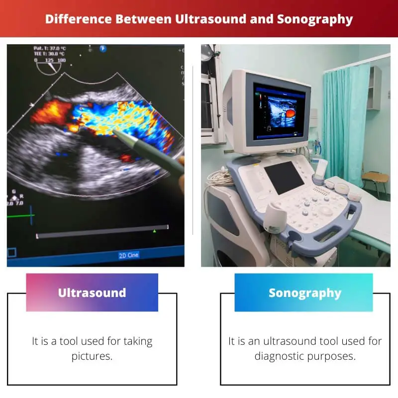
| Definition | It is a tool used for taking pictures. | It is an ultrasound tool used for diagnostic purposes. |
| Evolution | 1956 | The 1980s |
| Advantages | It is painless and doesn’t require injections. | It does not use radiation. |
| Disadvantages | If the depth is increased, then the lower frequency is required for optimal imaging. | It cannot penetrate bone. |
| Benefits | It is good for checking the health of the unborn baby. | It is safe and has no side effects. |
What is Ultrasound?
It is an imaging test. It uses waves for creating pictures of tissues, organs, and other structures of the body. The best part about ultrasound is they don’t use any radiation. It is done externally on the outside of the body.
People won’t feel any pain during this procedure and the only feeling they would get is the temperature of the translucent gel that is applied to the body for moving the transducer. Apart from that, the pain is virtually non-existent.
It is used in pregnant women to find the images of the baby in the uterus. This helps your healthcare provider to evaluate your baby’s growth so that they can monitor your pregnancy.
One of the various applications of ultrasound is you can get an image of the heart. It can reach the different parts of your body and points out the issues correctly.
Any flaws in the metal object can be easily detected. Gall bladder stones can be broken.
You should eat a low-fat dinner before you go for an ultrasound. Don’t eat or drink anything before the appointment as advised by the doctor. If you are required to take any medications, then drink only a small amount of water.
You should consume at least 8 glasses of water before your examination and your bladder should be full while they perform the examination.
Wear a two-piece outfit so that you don’t have to remove your entire outfit while they conduct the examination.

What is Sonography?
It is used for diagnosing and treating medical conditions. To become a sonographer, a person should study for an undergraduate sonography degree.
Some of the common pathways to this degree are science and nursing. Then complete a postgraduate degree in medical ultrasound.
Once you have gotten a postgraduate degree advance your career in this field with the postgraduate medical imaging degree. You can get your results soon, once they perform the test.
But in some cases, the images need to be analyzed it will take some time. In that case, the reports will be directly sent to the doctor.
The doctor will discuss the results later with you in the next appointment. Clinical training in sonography is difficult as the students will learn the technicalities of the job.
Since the clinical training is full-time, it can be stressful and strenuous as it is a real-world setting. But remember you are not alone; many people are available to help and guide you in the process.
When you work in this field, you will never be bored. Every day you will encounter something new. Because each day you will work with patients who approach you for different reasons.
Sometimes after the examination, your stomach might hurt for a few minutes, this is because of the pressure from the sonography wand.
Some people will fight this urge to pee many times because of the liquid that they consumed before the examination.

Main Differences Between Ultrasound and Sonography
- The definition of ultrasound is it is a tool used for taking pictures. On the other hand, the definition of sonography is it is an ultrasound tool used for diagnostic purposes.
- Ultrasound first came in the year 1956. On the other hand, sonography first came in the year 1980.
- The advantage of ultrasound is it is painless as it does not use injections. On the other hand, the advantage of sonography is it does not use any radiation.
- The downside of ultrasound is if the depth increases, then the lower frequency is required for optimal imaging. On the other hand, the downside of sonography is it cannot penetrate bone.
- The benefit of ultrasound is it is good for checking the health of an unborn baby. On the other hand, the benefit of sonography is it is safe and has no side effects.

- https://journals.sagepub.com/doi/abs/10.1016/j.carj.2016.08.002
- https://link.springer.com/chapter/10.1007/5584_2016_170
