The field of microbiology and bacteriology can not ply without microscopes. Science is becoming so advanced, and medicines are evolving more and more effectively only due to the detailed study of microorganisms.
Key Takeaways
- Bright-field microscopy uses transmitted light to illuminate the sample, creating a bright background, while dark-field microscopy scatters light around the specimen, resulting in a dark background.
- Darkfield microscopy excels at viewing transparent or translucent specimens, while bright-field microscopy is better suited for opaque samples.
- Bright-field microscopy is more commonly used and affordable, while dark-field microscopy is a specialized technique for specific applications.
Bright Field Microscope vs Dark Field Microscope
In bright field microscopy, the specimen is viewed under bright, uniform lighting and is suitable for observing stained specimens with high contrast. In dark field microscopy, the specimen is viewed under oblique, angled lighting and is better for visualizing unstained or transparent specimens.

The bright-field microscope is a popular and common microscope that does not cost much. It gets used to study specimens that are transparent and needs to get stained and fixed to be observed flawlessly.
This microscope is not always affordable due to its high cost; also, using it to examine it is not very handy.
Comparison Table
| Parameters of Comparison | Bright Field Microscope | Dark Field Microscope |
|---|---|---|
| Background Specimen Condition of specimen Resolution Expense Opaque disc Usability Studying minerals | In a bright field microscope, the background remains bright. | In a dark field microscope, the background is supposed to be dark. |
| Specimen | Its specimen is needed to be dark. | Its specimen is needed to be bright. |
| Condition of specimen | Here the specimen should be fixed and stained. | Here the specimen is not needed to be stained. |
| Resolution | Due to the staining of the sample, the resolution gets improved. | This microscope enhances the resolution without any staining. |
| Expense | These are not expensive. | This microscope is quite expensive. |
| Opaque disc | The opaque disc is not present in it. | Opaque disc exists in this microscope. |
| Usability | Using this microscope is quite easy. | Using this microscope is not easy. |
| Studying minerals | Studying minerals are not possible using this microscope. | Studying minerals, crystals, metals are possible with this microscope. |
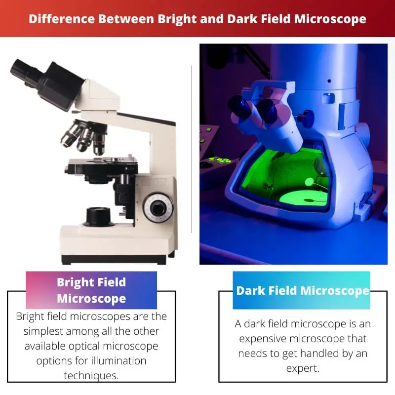
What is Bright Field Microscope?
In the field of microscopes, bright field microscopes are the simplest among all the other available optical microscope options for illumination techniques. Even an amateur can use this microscope and get the work done.
For viewing fixed specimens and live cells, this gets used as they are mostly transparent. And being easy to use, this type of microscope gets adopted to teach students.
However, as staining is a must condition here, the users should know the proper process of staining. Also, it requires powerful light for magnification, and the heat of such strong light can damage and even kill living sample organisms.

What is Dark Field Microscope?
A dark field microscope is an expensive microscope that needs to get handled by an expert. If you want to see specimens without staining them, this microscope must be utilized.
This can be used to identify thin bacteria, algae and fungi. Living cells can be observed using this without the chances of killing it with bright light.
The measurement can also be faulty. It also needs to get good care. The slide, stage, and even the light source must be free from dust.
To capture an accurate image, a longer time is needed. It also demands strong light, which can affect and harm the observer’s eyes.

Main Differences Between Bright and Dark Field Microscope
- The study specimens of bright field microscopes must be stained, but dark field microscope specimen does not require to be stained.
- Due to the staining, the resolution of the sample increases under a bright field microscope but under a dark field microscope, and the resolution improves without staining.
- https://link.springer.com/chapter/10.1007/978-3-540-70715-8_13
- https://ieeexplore.ieee.org/abstract/document/6588334/

A well-researched article overall, but the information on dark field microscopy seemed to be biased and made it sound more challenging to use than it actually is.
I see your point, but it’s important to highlight the challenges associated with dark-field microscopy for readers to have a balanced understanding.
The authors have done a good job covering the differences between bright-field and dark-field microscopy, but they could have added more information about the recent advances in these techniques for a more comprehensive understanding.
While the article does make some valid points, it failed to mention the limited field of view in a dark-field microscopy which could be seen as a disadvantage compared to a bright-field microscopy.
This article is on point and offers a very comprehensive and thorough analysis of the field of microbiology, and it’s essential for a deep understanding of the differences between bright-field and dark-field microscopy techniques.
The comparison table provided here perfectly summarizes the factors distinguishing these two microscopes. It’s an excellent, quick reference for those conducting research in this field.
I couldn’t agree more. The table is a fantastic addition to the article.
Yes, the comparison table adds value and makes it easy for readers to get a snapshot of these microscope types.
I have some reservations about the cost factor of bright-field microscopy mentioned in the article. One proper source to back this claim would have increased the credibility of the article.