Light microscopes had several drawbacks, leading to the electron microscope’s invention. Photon diffraction was the basis of light microscopes.
The concept of the electron microscope was born when it was discovered that it is also possible to achieve diffraction using electrons.
The wavelength of electrons is significantly less than that of photons because electrons have a significantly higher mass than protons.
As a result, electrons diffract less than photons. An electron microscope uses this principle to produce sharper images that can be magnified much more than a light microscope.
Two types of electron microscopes are most commonly used: scanning electron microscopes (SEM) and transmission electron microscopes (TEM).
Key Takeaways
- Scanning electron microscopes create images by scanning a focused electron beam on the surface of the specimen, providing detailed surface topography.
- Electron microscopes transmit electrons through a thin sample, generating high-resolution images of internal structures.
- Scanning electron microscopes offer a greater depth of field than transmission electron microscopes, allowing for three-dimensional imaging.
Scanning Electron Microscope vs Transmission Electron Microscope
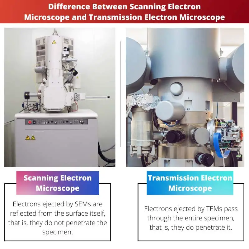
The difference between Scanning Electron Microscope and Transmission Electron Microscope is that scanning electron microscopes produce surface images by reflecting electrons from the specimen’s surface. In contrast, transmission electron microscopes produce an internal photo of the model by emitting electrons that cross through it.
An SEM ejects a refined and focused beam of electrons towards a sample. These electrons are captured after reflecting from the specimen’s surface.
A magnified image is created when electrons interact with the specimen’s surface. A magnified version of the specimen’s surface can be produced up to 2,000,000 times.
A TEM is an electron microscope that emits a broad beam of electrons. Electrons are captured after passing through this beam, penetrating the entire specimen.
Images of the specimen are thus obtained, showing its detailed internal structure. This image can be magnified up to 50,000,000 times.
Comparison Table
| Parameters of Comparison | Scanning Electron Microscope | Transmission Electron Microscope |
|---|---|---|
| Definition | Electrons ejected by TEMs pass through the entire specimen; that is, they do penetrate it. | Refined and focused electron beams are emitted by SEMs. |
| Image Produced | SEMs produce topographical or surface images of specimens. | TEMs provide detailed internal images of the specimen. |
| TEMs can produce 3D images. | SEMs produce topographical or surface pictures of specimens. | Electrons are emitted from TEMs in a broad beam. |
| Resolution | The images produced have a low resolution. | The images produced have a high resolution. |
| Image dimensions | Only 2D images can be produced by SEMs. | 3D images can be produced by TEMs. |
| Speed | SEMs are faster at processing images. | TEMs process images at a slower rate. |
| Magnification | SEMs have a magnification power of up to 2,000,000. | The magnification power of TEMs can reach 50,000,000 times. |
| Sample preparation | For magnification, SEMs do not require much sample preparation. | It is necessary to prepare the sample for magnification in TEMs. |
| Cost | The cost of operating SEMs is low. | TEMs have high operating costs. |
| Skills required | SEMs are easy to use and don’t require specialized skills to run. | TEMs are complicated devices that require some level of training to operate. |
What is a Scanning Electron Microscope?
A scanning electron microscope (SEM) employs electrons to illuminate a specimen and produce an enlarged image. It uses an electron gun to eject electrons from the model.
An electron beam is emitted from the gun. SEM emits electrons in a refined and focused beam.
As compared to a light microscope, which can also magnify substantially, SEM can create a higher-resolution image.
The electrons ejected by SEMs are reflected from the surface, so they do not penetrate the specimen. Using an SEM, samples can be imaged topographically or surface-wise.
SEMs emit refined and focused electron beams. The images they produce are of low resolution.
SEMs can produce only two-dimensional images. These devices process images more quickly.
The magnification powers of SEMs can reach up to 2,000,000. SEMs do not require much sample preparation before magnification.
They are relatively inexpensive to operate. Search engine marketing is easy to use and does not require specialized skills to work.

What is a Transmission Electron Microscope?
Another type of electron microscope is the transmission electron microscope.
Its resolution power is much greater than a photon projector because it creates images by projecting electrons rather than photons.
TEMs emit electron beams in the form of broad beams of electrons. The electrons ejected by TEMs pass through the entire specimen, so they do penetrate it.
These images give a detailed view of the internal structure of the specimen.
There is a high degree of resolution in the images produced. It is possible to create 3D images with TEMs.
The magnification power of TEMs reaches 50,000,000 times. TEMs process images more slowly.
In TEMs, the sample must be prepared for magnification. The operating costs of TEMs are high.
There is some training required to operate TEMs since they are complicated devices.

Main Differences Between Scanning Electron Microscope and Transmission Electron Microscope
- SEMs emit refined and focused electron beams reflected from the specimen’s surface. In contrast, TEMs emit electrons in a broad beam that passes through the entire specimen, thus penetrating it.
- SEMs produce low-resolution topographic or surface images of specimens, whereas TEMs produce high-resolution, detailed images of the specimen’s interior.
- The SEM processes images faster and has a magnification power of up to 2,000,000, while the TEM processes images at a slower rate and has a magnification power of up to 50,000,000.
- SEMs have a low operating cost, while TEMs have a high operating cost.
- An SEM is easy to use and does not require specific skills to run, while a TEM is a complex device requiring some training to operate.
- https://iopscience.iop.org/article/10.1088/0508-3443/6/11/304/meta
- https://link.springer.com/chapter/10.1007/978-1-4757-2519-3_1

This article is a bit difficult to follow, and some parts seem overly complicated. A simpler approach would have been more accessible.
The explanation provided is very informative and technically accurate. It adds a lot to the understanding of the topic.
I find the article’s explanation rather pedantic. It could have been simplified a bit for a wider audience.
A great, comprehensive article that gives a detailed overview of the topic. I would say, it’s in-depth enough to probably give a pretty solid understanding of SEMs and TEMs.
This article provides a fascinating insight into the world of electron microscopy. The thoroughness and depth of the explanation are truly commendable.
The article offers a very detailed explanation of the differences between SEMs and TEMs. It’s a great reference for scientists.
I absolutely agree with you. The writer did a great job explaining this complex topic in a very clear way.