Electrocardiograms (EKG) and echocardiograms (Echo) may sound similar, but they are not the same thing. An electrocardiogram (ECG) examines your heart’s electrical circuitry.
An echocardiogram (Echo) examines the mechanical system of the heart. The ECG and echocardiography are tests used to assess your heart’s general health.
There are several distinctions to be made between echocardiography and an electrocardiogram.
Key Takeaways
- ECG records electrical activity, while echocardiography creates visual images of the heart.
- ECG is quicker, less expensive, and non-invasive; echocardiography provides more detailed information about heart structures.
- ECG can detect arrhythmias and ischemia; echocardiography identifies valve abnormalities and evaluates cardiac function.
ECG vs Echocardiography
The difference between ECG and Echocardiography is that the former attaches electrodes to detect the results while the latter uses sound waves to detect the condition of the heart. Echocardiography is a moving image ultrasound of the heart that offers information on the anatomy and function of the heart. The ECG is a type of cardiac tracing that primarily offers information about the heart’s rhythm.

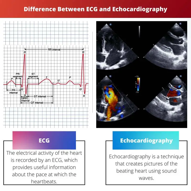
The heart’s electrical activity is recorded by an ECG, which provides useful information about the pace at which the heart beats, as well as the rhythm and regularity of the pulse.
An ECG is a rapid screening tool for arrhythmias, cardiac muscle damage following a heart attack, the status of any implanted device such as a pacemaker, and the detection of some congenital diseases and medication effects.
An ECG is also utilized as a part of a normal health checkup and as part of the workup performed before any major operation.
Echocardiography is a technique that creates pictures of the beating heart using sound waves. It employs 2-dimensional, 3-dimensional, and Doppler ultrasound techniques.
Transthoracic, transesophageal, and stress echocardiography are all options for echocardiography.
The doctor conducts the test by passing a transducer across the chest and connecting it to a monitor that records pictures of the heart. It takes no more than 10-15 minutes to complete the operation.
Comparison Table
| Parameters of Comparison | ECG | Echocardiography |
|---|---|---|
| Purpose | Recording of the heart’s electrical activity. | Creates pictures of the beating heart. |
| Uses | Used to detect damage of muscles in the heart or arrhythmias. | Detect the conditions of hearts. |
| Technique | Results are detected by attaching 12 patches to the chest, legs, and arms. | Results are detected by using a transducer over the chest area. |
| Form used | Electrodes are used to detect. | Uses sound waves to detect. |
| Form of result | Results are displayed on paper. | The images are shown on a computer. |
What is ECG?
An electrocardiogram (ECG) is a recording of the electrical activity of the heart. This is accomplished by placing non-painful electrodes on the skin’s surface that can capture this activity.
12 patches are applied to the chest, arms, and legs and are connected to a machine through cables. This equipment shows the electrical activity on a piece of paper, which may be interpreted.
The treatment takes less than 10 minutes and involves no electric shocks or bodily injury. During the activity, an ECG can be used to search for signs of cardiac stress.
An ECG is frequently used in conjunction with other tests to diagnose and monitor cardiac problems.
It can be used to look into symptoms, including chest discomfort, palpitations, dizziness, and shortness of breath that could indicate a heart condition.
An ECG can be used to diagnose, which includes Arrhythmias are heartbeats that are too slow, too fast, or irregular.
A build-up of fatty substances blocks or interrupts the heart’s blood flow, resulting in coronary heart disease.
Heart attacks occur when the heart’s blood supply is unexpectedly cut off. Cardiomyopathy is a condition in which the heart walls thicken or expand.
A series of ECGs can also be obtained over time to monitor someone diagnosed with a cardiac problem or receive medications that may affect the heart.

What is Echocardiography?
Echocardiography gives a wealth of information about the heart, including its size, shape, pumping capability, tissue damage location and degree, internal chambers, and valve function.
It is mostly used to assess the state of the cardiac muscle following a heart attack. It can identify infection in the sac around the heart as well as infection in the heart valves.
A color Doppler echocardiography can provide a detailed picture of the blood flow in the heart.
There are a variety of various echocardiograms that may be performed, which are transoesophageal echocardiography (TOE), in which a tiny probe is sent down the throat into the gullet and stomach, one may need to fast for several hours before this test.
Stress echocardiography is when an echocardiogram is performed during or shortly after a session on a treadmill or exercise bike activity or after receiving an injection of a medicine that causes the heart to work harder.
Contrast echocardiography, in which, before an echocardiogram, a harmless chemical called a contrast agent is injected into the circulation, this material shows up clearly on the scan and can assist in building a better image of the heart.
The sort of echocardiography you’ll have is determined by the heart disease being evaluated and the level of information required in the pictures.
Echocardiography is a painless, quick, and safe treatment.
The scan has no negative effects, however, the lubricating gel may feel chilly, and when the electrodes are withdrawn from over the skin after the test, one may feel some little pain.
Main Differences Between ECG and Echocardiography
- An ECG is mainly used for the recording of the heart’s electrical activity, whereas echocardiography creates pictures of the beating heart.
- An ECG examines a person’s heart rhythm, whereas an echocardiogram examines how the heart’s chambers and valves flow blood through the heart.
- In an ECG test, the results are detected by attaching 12 patches to the chest, legs, and arms, whereas in an Echocardiography test, results are detected by using a transducer over the chest area.
- An ECG uses electrodes to seek for abnormalities in the heart’s electrical impulses, whereas an echocardiogram uses ultrasonography to look for anomalies in the heart’s structure.
- The results are displayed on a paper after the ECG test, whereas in an echocardiography test, the results are displayed on the monitor during the test.
- https://www.jacc.org/doi/abs/10.1016/j.jcmg.2016.06.011
- https://academic.oup.com/eurjpc/article/21/6/774/5925783?login=true

I don’t think the article adequately addresses the possible risks and downsides of ECG and Echocardiography. More focus on the negative aspects would have given a more balanced viewpoint.
Thank you for this incredibly detailed comparison. I’ve always wondered about the differences between these two, and now I feel much more informed.
Finally, a clear and concise comparison between ECG and Echocardiography. I still get confused with these two. Thank you so much!
This article is a good example of how to take a dry topic and make it engaging to the reader. Well done!
This article was helpful and all, but I still think it could have included more real-world examples to better illustrate the differences between ECG and Echocardiography.
Very informative! Now I understand the key differences between ECG and Echocardiography. Thanks for the detailed and thoughtful explanation.