Benign tumours are a collective name for neoplasms of different origins, histological structures and localization. They can develop asymptomatically or present themselves with cough, hemoptysis, and shortness of breath.
In most cases, the treatment of such masses is surgical. It is believed that neoplasms arise as a result of genetic mutations and exposure to viruses, tobacco smoke, and chemical and radioactive substances. Tumours in the human body can have different structures depending on the cells underlying their formation.
Key Takeaways
- Hamartomas are benign growths composed of mature cells from the tissue where they develop.
- Teratomas are tumors containing multiple types of tissues derived from different germ layers.
- Teratomas are more aggressive and may become malignant, unlike hamartomas.

Hamartoma vs Teratoma
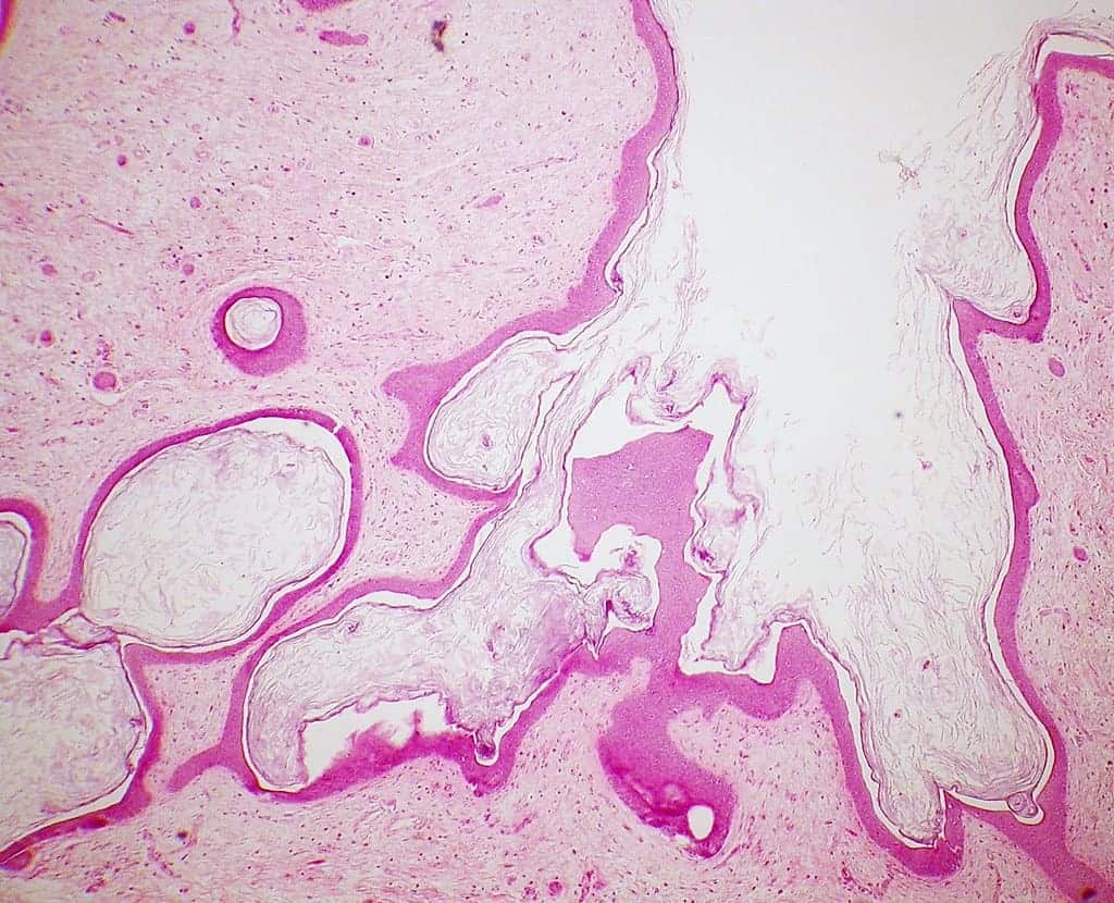
A teratoma is one of the most unusual tumours. Its structure is always heterogeneous and not peculiar to the anatomical area where it appeared. The neoplasm can contain several completely different types of tissues originating from one, two or three germ layers.
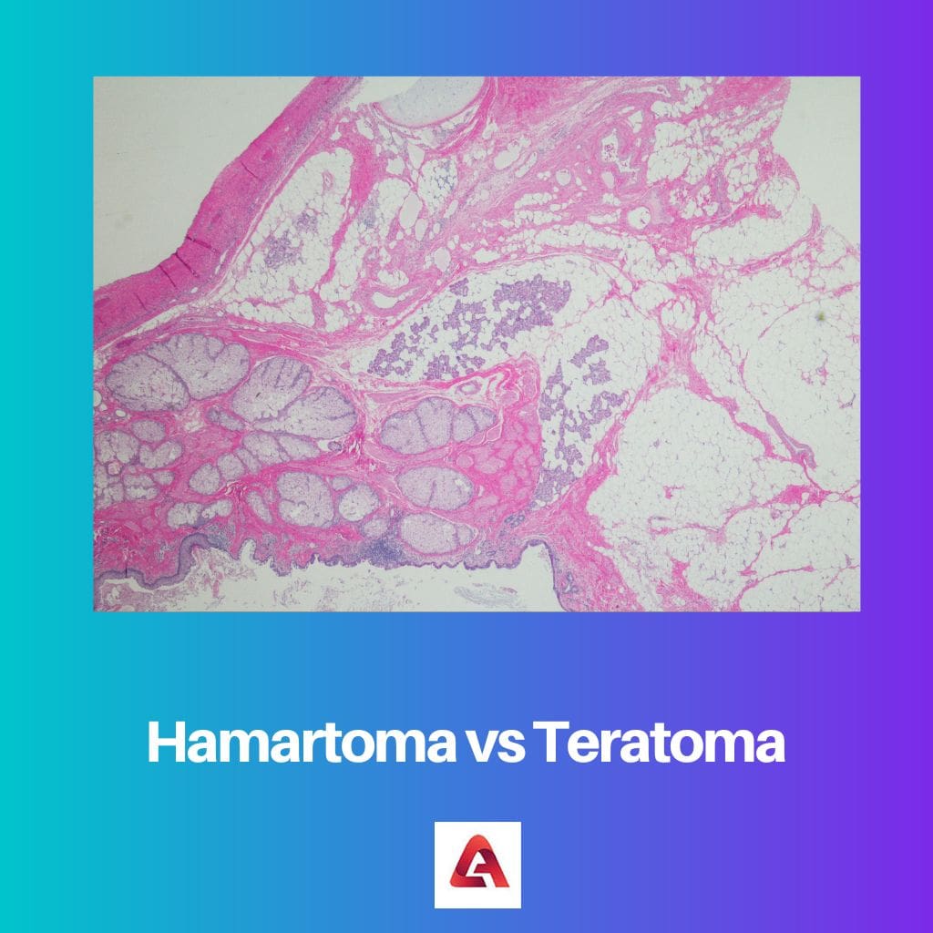
Hamartoma refers to tissue malformations and is an oversized component of an organ, including a vessel, due to growth abnormality. It looks like a node and is characterized by its benign form.
The difference between Hamartoma and Teratoma is that Hamartoma is a benign mass that forms in the lung during the stage of intrauterine development. On the other hand, Teratoma is a tumour that grows from the cells of an embryo. It develops even before a person is born, and its manifestations are possible at any age.
Hamartoma is a benign neoplasm with predominant localization in the lungs. Retinal hamartoma and hepatic hamartoma are very rare. A benign tumour is a slow-growing neoplasm without sprouting into neighbouring tissues. Such tumours don’t metastasize or malign. The severity of symptoms depends on size and localization.
A teratoma is a tumour growing from the cells of an embryo. The main reason for the development of the tumour is a disruption of the normal development of the embryonic tissues. This phenomenon leads to the teratoma containing the rudiments of several organs, which are not typical for this anatomical structure.
Comparison Table
| Parameters of Comparison | Hamartoma | Teratoma |
|---|---|---|
| Definition | Hamartoma can be defined as a mass of cells growing in an organ | Teratoma is a growth that forms from all three germ layers and may even contain hair and teeth in itself |
| Causes | Cowden syndrome or tuberous sclerosis | Due to genetic mutations that cause abnormal cell and tissue development |
| Diagnosis | X-ray, tomography scan | Ultrasound, X-rays, and CT scans |
| Symptoms | Pain, heart failure, seizures | Pain, unusual bleeding |
| Treatment | Knife radiation surgery | Can be removed by surgery |
What is Hamartoma?
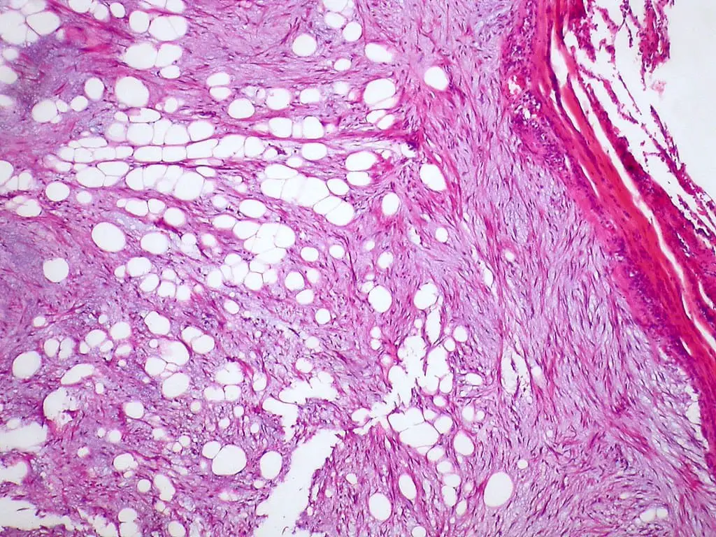
A hamartoma is a non-malignant type of tumour. The cells in this type of tumour resemble normal cells. However, unlike normal, healthy cells, hamartoma cells are disorganized and have no function for the affected organ. A hamartoma may consist of one or more cell types.
Hamartomas can develop anywhere in the body, although they are more commonly found in the lungs, digestive tract (especially the colon), mammary gland, and brain. The type of cells found in a hamartoma will depend on the affected body area. Although hamartomas are not considered cancer, they can still cause damage by squeezing nearby structures such as other organs, nerves, or blood vessels.
In addition to parenchymatous lung cells, a hamartoma may contain cartilaginous, connective, and fatty components. The shape is round, with sizes ranging from five to fifty millimetres or more.

What is Teratoma?
A teratoma is formed due to the abnormal development of embryonic tissues. Its growth begins even before the birth of the child. It is formed from germ cells that can grow into any human body tissue.
Teratomas are undifferentiated germ cell neoplasms of embryonic origin. Their development is based on the violation of differentiation and migration of primary germ cells. Therefore, teratomas are localized in the glands. Tumours can also be located in the mediastinum, brain, sacrococcygeal area, and other anatomical regions. They are more frequently diagnosed in childhood or adolescence and less frequently in adults or during intrauterine development. In the latter case, a large teratoma can hinder the development of the fetus and prevent natural childbirth.
According to statistics, embryomas account for 24% to 36% of the total number of neoplasms in children and 2.7-7% in adults. They occur in boys and girls. Some neoplasms appear in the first years of a child’s life, while others “announce themselves” for the first time during the hormonal reorganization of the body.

Main Differences Between Hamartoma and Teratoma
Hamartoma
- A benign mass of disorganized tissue.
- The cause of hamartoma is the PTEN genetic mutation.
- A focal growth resembling a neoplasm but resulting from the abnormal development of an organ.
- A hamartoma is a predominantly benign local cell mass resembling a local tissue neoplasm, but due to a proliferation of multiple aberrant cells, based on a systemic genetic disease rather than growth originating from a single mutated cell, which defines a benign neoplasm/tumour.
- The choice of treatment method depends on where the hamartoma is located.
Teratoma
- A benign or malignant tumour arises from germ cells and consists of different types of tissue, such as skin, hair, or muscle.
- A tumour is sometimes found in newborn children that consists of a heterogeneous mixture of tissues, like bone, cartilage, and muscle.
- A tumour consists of a mixture of tissues that are not normally found in a given location.
- Teratomas are removed surgically.
- Teratoma has a mixed structure.
- https://www.sciencedirect.com/science/article/abs/pii/S0165587602003762
- https://www.ncbi.nlm.nih.gov/pmc/articles/PMC5426149/
