The peritoneal cavity begins and continues inside the abdominal cavity until it eventually transforms into the pelvic cavity. There are no organs in it but does have a thin layer of peritoneal fluid. The peritoneal surfaces are lubricated by this fluid, enabling the membranes to slide over one another.
Two separate layers make up the peritoneum. The visceral and parietal peritoneum are two of the layers that flow into one another continuously. This article examines these two levels in further detail, highlighting the variations that exist between them.
Key Takeaways
- The parietal peritoneum lines the abdominal cavity wall, while the visceral peritoneum covers the internal organs within the cavity.
- The parietal peritoneum is sensitive to pain, pressure, and temperature, whereas the visceral peritoneum is not directly sensitive to these stimuli.
- The parietal peritoneum derives from the somatic mesoderm, while the visceral peritoneum originates from the splanchnic mesoderm.

Parietal Peritoneum vs Visceral Peritoneum
The parietal peritoneum is the outer layer of the peritoneum that lines the abdominal wall and the undersurface of the diaphragm. It is attached to the abdominal wall and forms a continuous lining. The visceral peritoneum is the inner layer of the peritoneum that covers the abdominal organs.
The parietal peritoneum gets the equivalent somatic nerve supply as the area of the abdominal wall that it borders, making pain from it well localized. The parietal peritoneum is temperature-sensitive, with pressure, discomfort, and laceration.
On the other hand, the abdominal viscera are covered by the invaginated visceral peritoneum. It comes from the embryo’s splanchnic mesoderm.
The same autonomic nervous system supplies the viscera it covers as the visceral peritoneum. The visceral peritoneum only responds to chemical irritation and stretch, unlike the parietal peritoneum, which has poor localization of pain.
Comparison Table
| Parameters of Comparison | Parietal Peritoneum | Visceral Peritoneum |
|---|---|---|
| Definition | The membrane covers the interior surface of the diaphragm and abdominopelvic wall. | The membrane covers most of the abdominal organs by folding back or turning inside out. |
| Sensitiveness | Pressure, pain. | Specific chemical exposure. |
| Membrance function | Interior lines between the abdomen and the pelvis. | Most abdominal organs are lined inside the abdominal cavity. |
| Blood and nerve supply | Areas are connected along the abdominal wall lining. | Areas are connected along protected organs. |
| Pain experience | Specific area becomes severe. | Presence of dull pain in the general area. |
What is Parietal Peritoneum?
The term “parietal peritoneum” refers to the peritoneum’s outer layer, which covers the diaphragm as well as the walls of the abdomen and pelvis. It is an embryological offshoot of the mesoderm, consisting of a single layer of mesothelial cells attached to fibrous tissue.
A thin membrane called the peritoneum lines the abdominopelvic cavity. It has two layers: the inner visceral layer, which encircles the abdominal organs, and the outermost parietal layer, also known as the parietal peritoneum, which covers the belly and pelvis.
A possible space between these layers includes a tiny quantity of serous fluid (about 50–100 mL), which is made up of water, electrolytes, and immune cells (e.g., white blood cells). This liquid is a type of protection and lubrication between the layers.
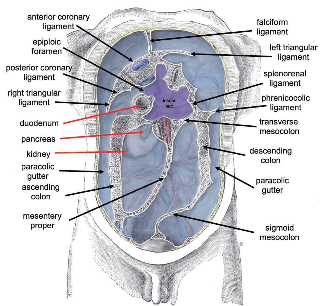
Depending on whether the peritoneum completely or only partially covers an organ, it is classified as intraperitoneal or retroperitoneal. Organs like the liver, stomach and transverse colon are examples of intraperitoneal structures or organs because they are entirely encased in the visceral peritoneum.
The parietal peritoneum, more particularly, solely covers the anterior wall. Depending on where they were during development, these organs are further divided into primary and secondary retroperitoneal organs. The adrenal glands, kidneys, ureters, abdominal aorta, inferior vena cava, and corresponding branches are among the primary retroperitoneal organs that develop and stay outside the peritoneal cavity.
The pancreas, the ascending and descending colon, and the duodenum distal portion are secondary retroperitoneal organs, which initially develop intraperitoneally and transform into retroperitoneal structures throughout development.

What is Visceral Peritoneum?
The membrane that folds back or turns inside out to cover the majority of the abdominal organs is known as the visceral peritoneum. During early embryonic stages, it is also formed from somatic mesoderm.
The same autonomic nerve that connects to the organ it covers also supplies the visceral peritoneum with blood and nerves. The visceral peritoneum’s nerves are more sensitive to stretching or exposure to specific substances than the parietal peritoneum’s.
Pain from the visceral peritoneum affects skin regions supplied by the same neurological sensory fibres that flow into the abdominal organs. Pain that originates in the visceral peritoneum is frequently subtle and difficult to pinpoint. It appears as a generalized area of pain.
Main Differences Between Parietal Peritoneum and Visceral Peritoneum
Parietal Peritoneum
- The peritoneum’s outer layer, known as the parietal peritoneum, covers the diaphragm as well as the abdominal and pelvic walls.
- The parietal peritoneum protects and supports the nerves, blood arteries, and lymphatic vessels that supply the belly and pelvis.
- The abdominal wall that borders the parietal peritoneum shares the same innervation.
- The walls of the abdominal and pelvic cavities are lined with the parietal peritoneum, which is found in the abdomen.
- The parietal peritoneum, an embryological descendant of the mesoderm, is made up of a single layer of mesothelial cells attached to fibrous tissue.
Visceral Peritoneum
- As it wraps around your organs, the visceral peritoneum folds up on itself, forming pockets.
- When your digestive organs are swollen from food or gas, it feels stretched.
- It detects temperature, discomfort, and localized pressure.
- The same autonomic nerve that connects to the organ it covers also supplies the visceral peritoneum with blood and nerves.
- Pain occurring in the visceral peritoneum is subtle and difficult to pinpoint
