There are a ton of confusions encompassing biometric innovation. One of the most essential is the disarray about iris acknowledgment and retinal outputs. An astounding number of individuals utilize these terms reciprocally.
While both include portions of the eye, they are altogether different. Since it’s precise, stable, harmless, and quick, iris acknowledgment is utilized broadly across the globe.
Key Takeaways
- The retina is a light-sensitive layer at the back of the eye that converts light signals into neural impulses. At the same time, the iris is a pigmented, muscular structure that controls the amount of light entering the eye.
- Retinal cells contain photoreceptors, which enable vision by detecting light and generating electrical signals, while the iris adjusts pupil size to regulate light exposure.
- The retina plays a crucial role in the image formation and visual perception, while the iris contributes to eye color and helps maintain optimal lighting conditions for vision.
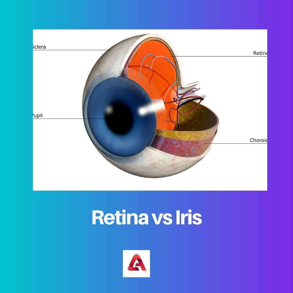
Retina vs Iris
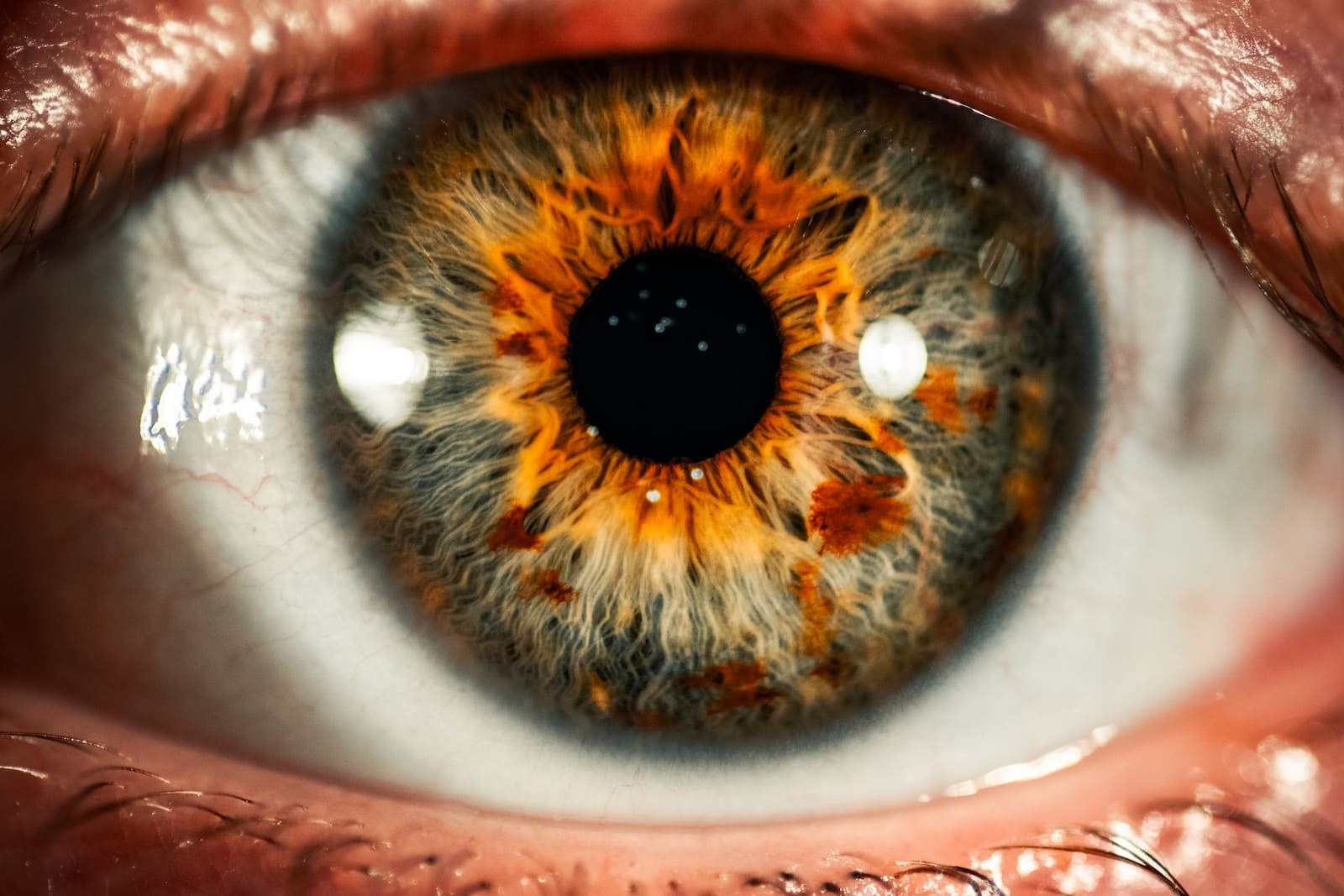
The difference between retina and iris is that retina is not visible properly. On the other hand, iris is the part of the eye that comes off as colorful. The capacity to separate between retinal checking and iris acknowledgment is fundamental if you are thinking about putting resources into biometrics and anticipate an exceptional yield on speculation.
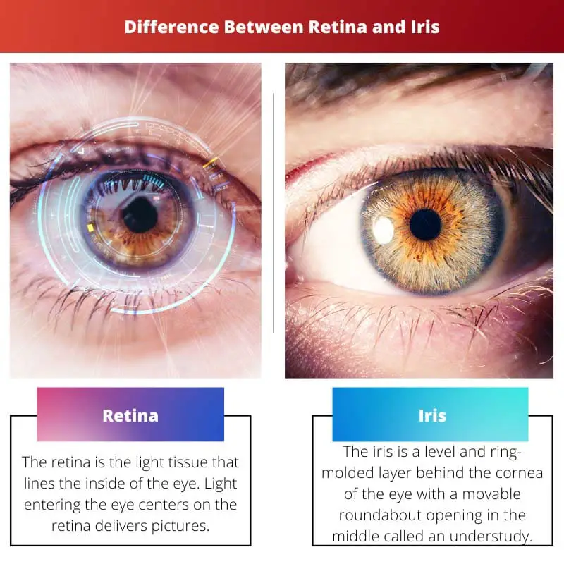
The retina is the light tissue that lines the inside of the eye. Light entering the eye centers on the retina delivers pictures. The subsequent light signals are then communicated from this light-touchy tissue, through the optic nerve to the cerebrum.
This is the essential pathway of vision. On the off chance that the retina tumbles off within the rear of the eye (retinal separation), vision is lost. On the off chance that the macula (the focal part of the retina) isn’t smooth, vision is writhed.
The iris is a level and ring-molded layer behind the cornea of the eye with a movable roundabout opening in the middle called an understudy. This is the construction that gives a singular eye tone.
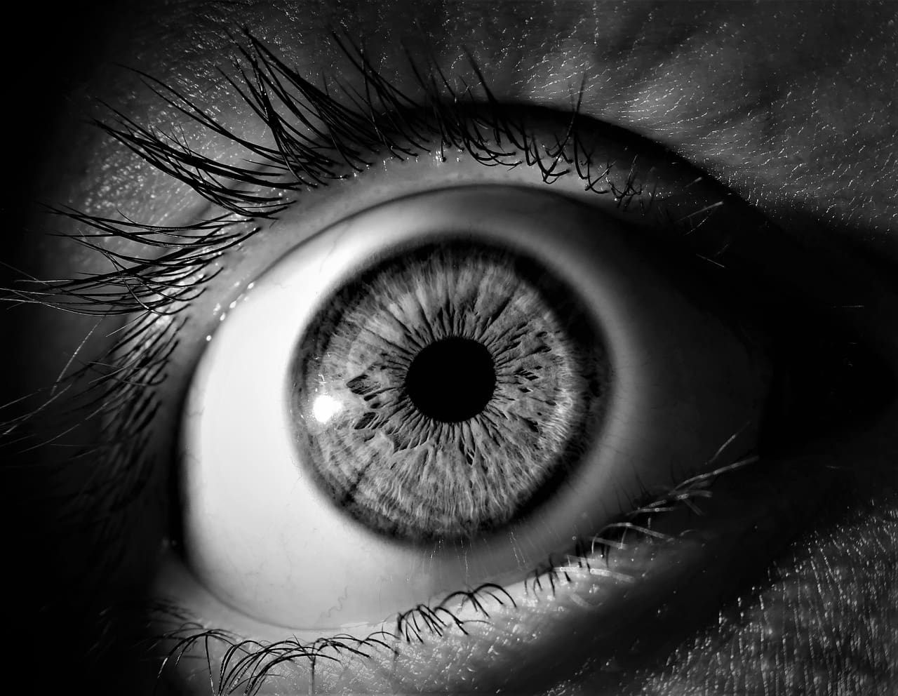
Along with the student, the iris is answerable for controlling how much light that gets into the eye. To an extreme or too minimal light can hamper vision.
The solid iris moves to shrivel the student assuming there is a lot of light and enlarge it on the off chance that it isn’t sufficient.
Comparison Table
| Parameters of Comparison | Retina | Iris |
|---|---|---|
| Impact | Disease can impair retinal scanning precision. | Iris remains relatively unchanged. |
| Distance | Retinal scanning necessitates bringing the eye very near to a lense. | Iris recognition can be done from a range. |
| Acknowledgment | Retinal scanning as an advertising biometric mode is not broadly acknowledged. | Iris recognition as an advertising biometric mode is broadly acknowledged. |
| Intrusiveness | Retinal scanning is regarded as intrusive. | Iris recognition is non-invasive. |
| Location | The retina is certainly not an apparent piece of the eye. | The iris is the hued piece of your eye encompassing the understudy. |
What is Retina?
The retina is an overlaid tissue. It has 3 distinct practical layers. The peripheral layer is the photoreceptor layer, composed of bars and cones.
These phones change the light energy into electrical signals that are handled and then shipped off the cerebrum where vision is finished. Geographically, the focal point of the retina is known as the macula.
It is here that focal vision and nitty-gritty vision happen. Both the cornea and our normal focal point shine light onto the retina. Sometimes patients require contact focal points and glasses to assist with this interaction.
These forms and the glassy body that fills the focal point of the eye should be clear for the light to get to the retina.
The most well-known issue slowing down this cycle is a waterfall, an opacification of the focal point. In ordinary circumstances, the retina should be good so that we might do well.
Despite the thickest and best glasses, if this tissue isn’t sound, we won’t see. In the natural eye, its veins support the retina that feeds the inward layers and the choroid that feeds the external layers of the retina.
The internal veins are the ones that are burdened by diabetic retinopathy. Vascular impediments, both vein and impediments, can prompt loss of ordinary blood supply to the retina and lead to visual impairment.
The choroidal veins are the wellspring of liquid and drain in wet age-related macular degeneration (AMD).
What is Iris?
The iris is the piece of the eye that makes up your eye tone. A roundabout muscle with an opening in the center, the iris agreements and extends to control how much light that gets into the student.
The iris is made primarily of connective tissue and smooth muscle filaments. Despite a typical conviction, real change in the shade of the iris seldom occurs.
While the shade of an eye might seem to change, this is normally because of lighting changes or insight based on neighboring tones. Various problems, infections, and other ailments can influence the iris, and, likewise, the visual framework overall.
Checking for the strength of the iris as well as appropriate pupillary reflexes is a significant piece of care; not exclusively are these expected to analyze conditions, they additionally permit specialists to know whether this piece of the eye is working regularly.
Fortunately, eye-trained professionals (ophthalmologists) and optometrists have various tests they can utilize.
The iris sits in the uveal lot, which is the eye’s center layer. The iris lies before the focal point and behind the cornea. The iris controls how much light that enters the eye by opening and shutting the understudy.
The iris utilizes muscles to change the size of the understudy. These muscles can handle how much light enters the eye by making the student bigger (expanded) or more modest (tightened).
Main Differences Between Retina and Iris
- Retinal examining precision can be impacted by illness; the fine surface of the iris remains surprisingly steady.
- Iris acknowledgment resembles taking a typical photo of an individual and can be performed in a good way; though retinal examining requires the eye to be carried extremely near an eyepiece.
- Iris’s acknowledgment is all the more broadly acknowledged as a business biometric methodology than retinal examining.
- While both are contactless, retinal filtering is viewed as intrusive because it radiates apparent light into the eye through iris acknowledgment involves a computerized photo for recognizable proof, and is painless.
- The iris is the colored component of your eye that surrounds the pupil, whereas the retina is not noticeable.