Humans are very dependent on their ability to see. The sense of sight not only alerts us to threats to our survival but also enriches our existence with descriptions of different colors and other vitals of the world.
From the retinal receptors to the cerebrum’s primary visual cortex, the visual system consists of the eye and a long pathway of neural connections.
An image of the external world is formed in our brain by processing the pattern of excitations within the retina. Several regions inside the occipital lobe interact to form this central representation that simultaneously processes different aspects of the input.
Key Takeaways
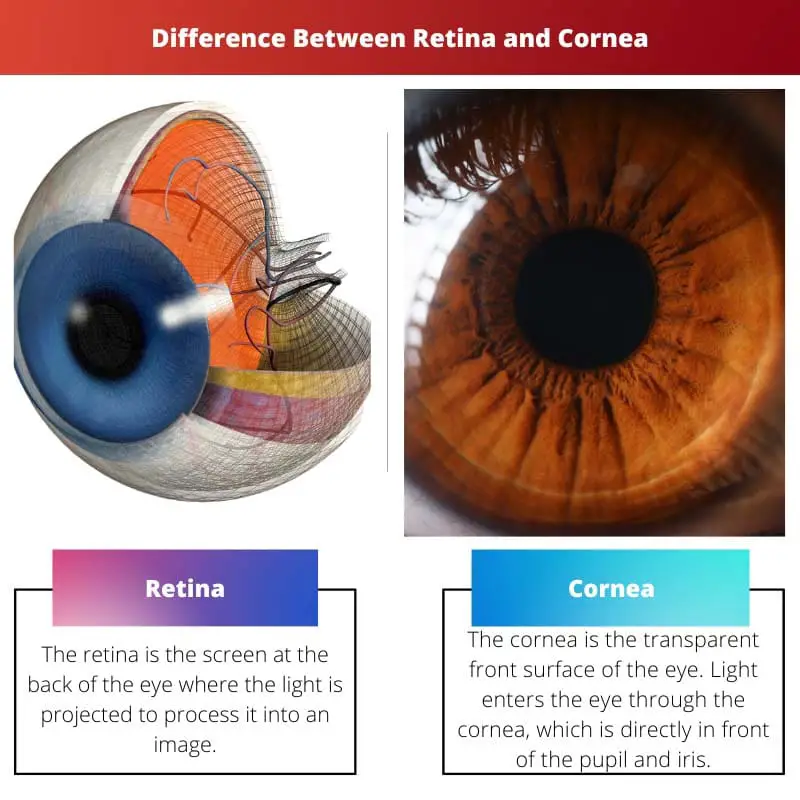
- The retina is a light-sensitive layer of tissue located at the back of the eye, responsible for converting light into electrical signals that the brain interprets as visual images. At the same time, the cornea is the clear, dome-shaped outer layer of the eye that helps focus light entering the eye.
- Retinal disorders, such as macular degeneration or retinal detachment, can result in vision loss or blindness. At the same time, corneal issues, like abrasions or infections, can cause pain, blurred vision, or light sensitivity.
- Treatment for retinal conditions may involve medications, laser therapy, or surgery, depending on the severity and underlying cause. In contrast, corneal issues may require eye drops, antibiotics, or corneal transplants in more severe cases.
Retina vs Cornea
The projection of light to form an image in an eye is made at the retina. The retina is made up of millions of cells. The size of the retina is 1094 square mm. Images formed by the retina are sent to the brain, where the brain allows the eyes to see the image. The transparent part of the eye is the cornea. The size of the cornea is 11.5 mm.
Millions of cells that are sensitive to light make up the retina, along with other nerve cells responsible for receiving and processing visual information.
Through your optic nerve, your retina sends these images to your brain, allowing you to see. This is the membrane that lines the back of the eyeball and provides the sense of touch.
Multiple layers make up the retina, including specialized cells known as photoreceptors.
The cornea is the transparent front surface of the eye. Light enters the eye through the cornea directly in front of the pupil and iris.
When looking at the cornea from the front, it looks wider than tall. It is because the sclera slightly overlaps the front and back surfaces of the anterior cornea.
Comparison Table
| Parameters of Comparison | Retina | Cornea |
|---|---|---|
| Colour | Orange Glow | Clear |
| Location | At the back of the eyes | In front of the iris and pupil |
| Importance | Processes the light into an image | Directs the light towards the retina |
| Size | 1,094 square mm | 11.5 mm |
| Disease | Retinitis pigmentosa | Keratitis |
What is Retina?
The retina is the screen at the back of the eye where the light is projected to process it into an image. Millions of cells that are sensitive to light make up the retina, along with other nerve cells responsible for receiving and processing visual information.
Through your optic nerve, your retina sends these images to your brain, allowing you to see. This is the membrane that lines the back of the eyeball and provides the sense of touch.
Multiple layers make up the retina, including specialized cells known as photoreceptors. These tiny receptors are of two types:
- Cones: These cones are located at the macula, the retina’s centre. It is these cells that allow for detailed vision and color perception. Reading and driving are made possible because of the macula, which provides high-definition vision.
- Rods: The outer retinal cells are denser than the inner ones. These cells are called rods. You can see in low lighting with these cells, which are used in peripheral vision.
There are various types of retinal diseases. Some of them are listed below –
- Retinal tear
- Retinal detachment
- Diabetic retinopathy
- Epiretinal membrane
- Macular hole
- Macular degeneration
- Retinitis pigmentosa

What is Cornea?
The cornea is the transparent front surface of the eye. Light enters the eye through the cornea directly in front of the pupil and iris. When looking at the cornea from the front, it looks wider than tall.
It is because the sclera slightly overlaps the front and back surfaces of the anterior cornea.
Patients who suffer from farsightedness, nearsightedness, astigmatism, or other eye disorders can benefit from corneal surgery. In addition to corneal transplants, Descemet’s Stripping Automated Endothelial Keratomileusis (DSAEK) is a relatively new procedure.
For those with damaged or cloudy lenses, donor corneas can be used to restore their vision. The newer technique replaces the cornea only partially, allowing for faster healing. Some of the most common corneal diseases are listed below:
- Keratitis: It refers to the reddening of the cornea. The inflammation due to bad-quality contact lenses leads to keratitis.
- Dry eye refers to when our eyes don’t secrete enough fluids to keep themselves wet, thus leading to visual problems and inflammation.
- Corneal dystrophies: This refers to the cloudy vision of the eyes due to accumulated waste near the cornea. If not treated, this waste could lead to a permanent cloudy vision.

Main Differences Between Retina and Cornea
- The retina is located at the back of the eyes, whereas the cornea is located in front of the iris and the pupil.
- The color of the retina has an orange glow and is translucent, whereas the color of the cornea is clear.
- The retina processes the light into an image, whereas the cornea directs that light towards the retina.
- The size of the retina is 1094 square mm, whereas the size of the cornea is 11.5 mm.
- The retinal disease is termed retinitis, whereas the corneal disease is termed keratitis.
- https://www.pnas.org/doi/abs/10.1073/pnas.1516259112
- https://www.liebertpub.com/doi/full/10.1089/hum.2020.070
