MRI (Magnetic Resonance Imaging) captures detailed images of anatomical structures using magnetic fields and radio waves. fMRI (functional Magnetic Resonance Imaging) measures blood flow changes in the brain, providing insights into neural activity during specific tasks or stimuli, making it suitable for studying brain function and connectivity.
Key Takeaways
- MRI (Magnetic Resonance Imaging) and fMRI (Functional Magnetic Resonance Imaging) are medical imaging tests. Still, fMRI is used to observe brain activity, while MRI is used to diagnose structural abnormalities.
- MRI uses strong magnetic fields and radio waves to produce detailed images of the inside of the body, while fMRI measures changes in blood flow to brain areas in response to different stimuli.
- While MRI is used to diagnose various conditions such as tumors and internal injuries, fMRI is used mainly for research purposes to study brain function.
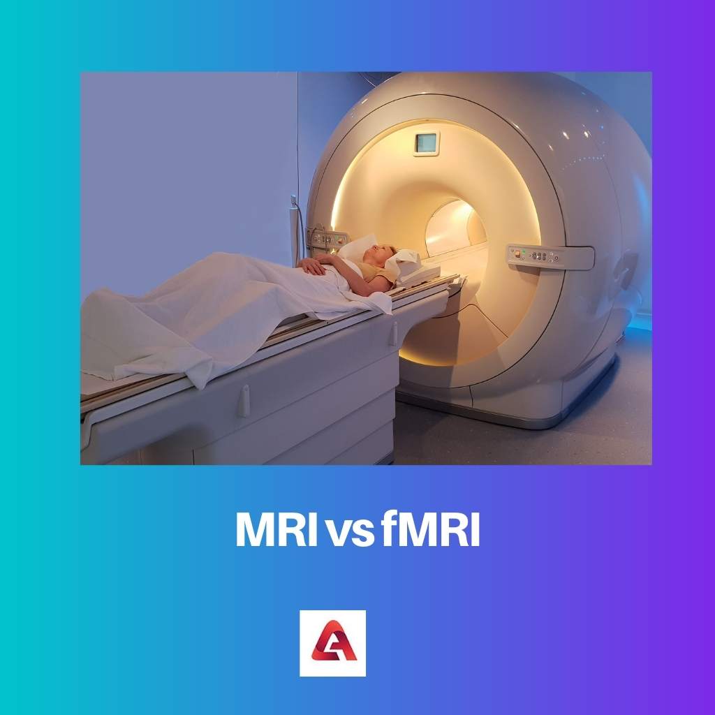
MRI vs fMRI
The brain’s anatomical structure can be determined through a machine called MRI. Problems related to the brain can be scanned through an MRI machine. The brain’s metabolic function can be determined through a machine called fMRI. fMRI is used only in experimental processes yet. fMRI machines are costly. Extra hardware and software are required for fMRI to work.
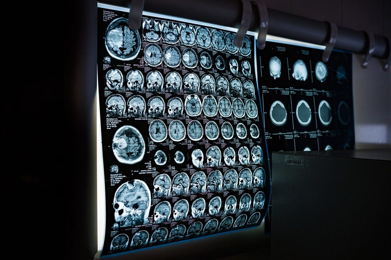
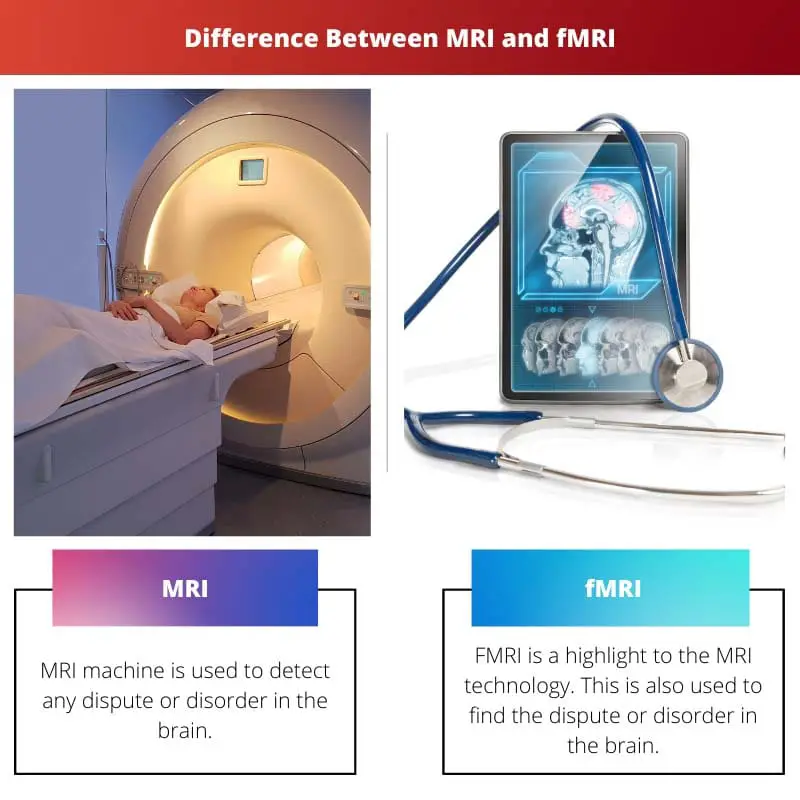
MRI machine is used to detect any dysfunctionality or disorder in the brain. The CT scan sometimes fails to detect the problem, so MRI was introduced to overcome the problem.
fMRI is a highlight of the MRI technology. This is also used to find the dispute or disorder in the brain. The full form of fMRI is Functional Magnetic Resonance Investigation.
Comparison Table
| Feature | MRI | fMRI |
|---|---|---|
| Full Name | Magnetic Resonance Imaging | Functional Magnetic Resonance Imaging |
| Purpose | Creates detailed anatomical images of the body’s interior | Measures brain activity by tracking blood flow changes |
| Information Provided | Structure of organs, tissues, bones, and abnormalities | Active areas of the brain during specific tasks |
| Applications | Diagnosis of various medical conditions like tumors, injuries, and abnormalities | Studying brain function in language, memory, decision-making, and mental health |
| Body Area Examined | Can be used for various body parts like brain, spine, knees, abdomen, etc. | Primarily focused on the brain |
| Procedure | Similar for both, involving a strong magnetic field and radio waves within a scanner | May require performing specific tasks or resting while in the scanner |
| Time | Can vary depending on the area examined (typically 30-60 minutes) | Slightly longer than MRI due to recording activity over time |
| Cost | Generally more expensive than X-rays or CT scans, but cost can vary depending on facility and region | Typically more expensive than a standard MRI |
What is MRI?
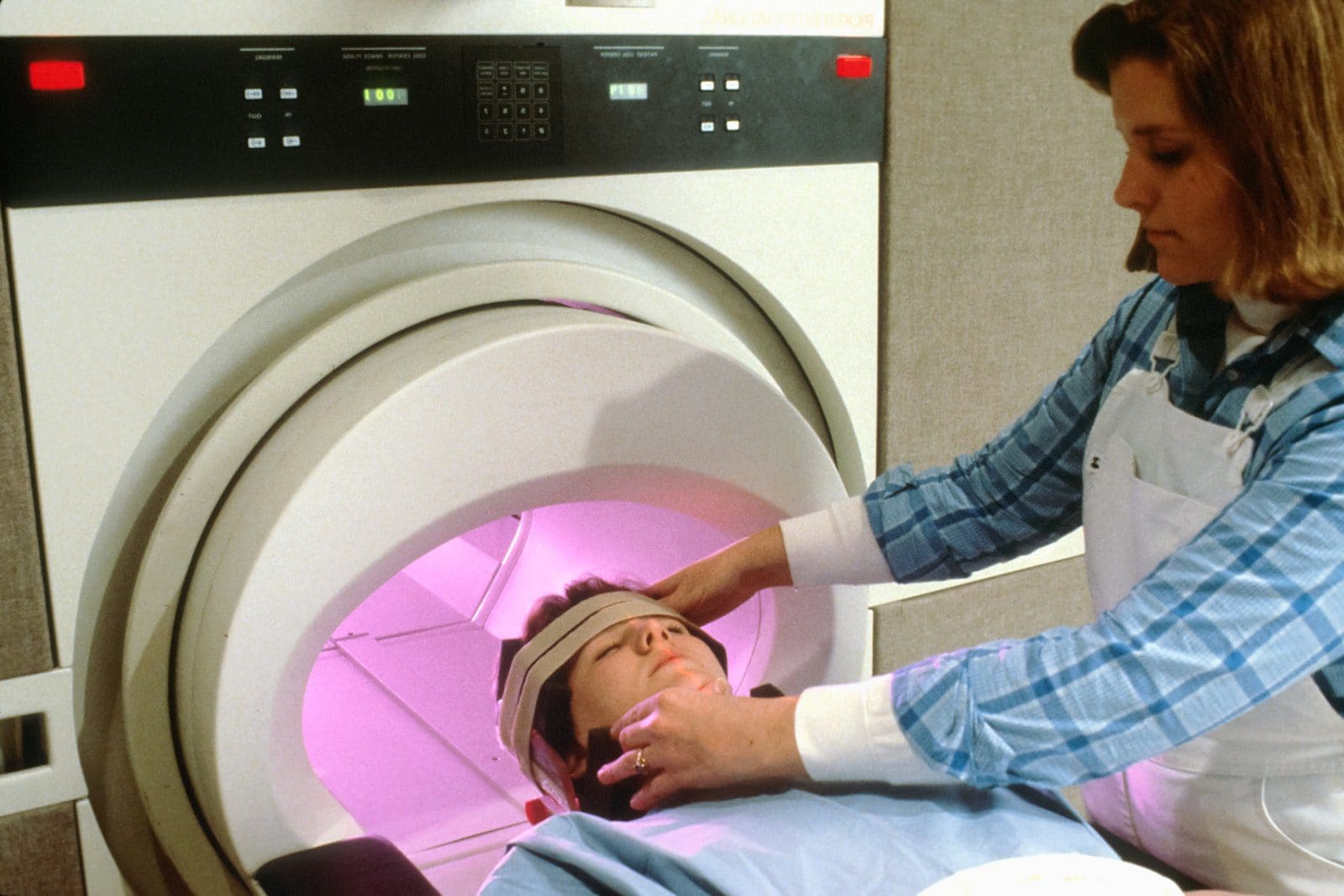
Magnetic Resonance Imaging (MRI) is a sophisticated medical imaging technique that utilizes powerful magnets and radio waves to generate detailed images of the internal structures of the human body. It has become a vital tool in the field of diagnostic medicine due to its non-invasive nature and ability to provide high-resolution images of soft tissues.
Working Principle
Magnetic Fields
At the core of MRI is the interaction between magnetic fields and the body’s water molecules. When a patient is placed in a strong magnetic field (typically generated by a superconducting magnet), the hydrogen protons in water align themselves with this magnetic field.
Radiofrequency Pulses
Radiofrequency pulses are then applied, causing the hydrogen protons to temporarily move out of alignment. When these pulses are turned off, the protons return to their original alignment, releasing energy in the form of radiofrequency signals.
Signal Detection
A receiver coil picks up these signals, and sophisticated computer algorithms convert them into detailed images. The strength and duration of the signals provide information about the density and distribution of water molecules in different tissues, enabling the creation of highly detailed anatomical images.
Types of MRI
T1-weighted and T2-weighted Imaging
Different types of MRI sequences highlight specific tissue characteristics. T1-weighted images emphasize the density of certain tissues, while T2-weighted images accentuate differences in water content.
Functional MRI (fMRI)
Functional MRI is used to assess brain activity by detecting changes in blood flow. It has applications in neuroscience and helps researchers understand the brain’s functional regions.
Diffusion-weighted Imaging (DWI)
DWI measures the random motion of water molecules in tissues, providing valuable information about cell integrity and detecting conditions like strokes or tumors.
Clinical Applications
Neuroimaging
MRI is widely employed for brain imaging, aiding in the diagnosis of neurological disorders such as tumors, multiple sclerosis, and vascular abnormalities.
Musculoskeletal Imaging
In orthopedics, MRI helps assess soft tissues, ligaments, and joints, providing valuable information for diagnosing conditions like torn ligaments, arthritis, and disc herniation.
Cardiovascular Imaging
MRI plays a crucial role in cardiovascular medicine, enabling detailed imaging of the heart and blood vessels, helping diagnose conditions such as heart attacks and aneurysms.
Oncology
In oncology, MRI is instrumental for detecting and staging tumors in various parts of the body, guiding treatment planning.
Advantages and Limitations
Advantages
- Non-ionizing radiation: Unlike X-rays, MRI does not use ionizing radiation, making it safer for repeated use.
- High soft tissue contrast: MRI excels in visualizing soft tissues, making it ideal for certain diagnostic scenarios.
Limitations
- Cost and accessibility: MRI machines are expensive to acquire and maintain, limiting their availability in some regions.
- Contraindications: Patients with certain metallic implants or devices may be restricted from undergoing an MRI.
What is fMRI?
Functional Magnetic Resonance Imaging (fMRI) is a non-invasive neuroimaging technique that allows researchers to observe and measure brain activity by detecting changes in blood flow. It has become a powerful tool in the field of neuroscience, enabling the investigation of various cognitive processes and functions.
How fMRI Works
1. Blood Oxygenation Level Dependent (BOLD) Signal
fMRI relies on the BOLD effect, which measures the magnetic properties of hemoglobin. When neurons are active, they require more oxygen, leading to increased blood flow to the active brain regions. The BOLD signal captures the changes in blood oxygenation, providing a proxy for neural activity.
2. Magnetic Resonance Imaging (MRI)
fMRI uses MRI technology, which involves exposing the brain to a strong magnetic field and radio waves. The interaction between these elements produces detailed images of brain structures. Functional MRI extends this by capturing changes in the MRI signal over time.
Applications of fMRI
1. Cognitive Neuroscience
fMRI is widely used to study cognitive processes such as memory, attention, language, and perception. By correlating brain activity with specific tasks, researchers gain insights into how different regions contribute to cognitive functions.
2. Clinical Applications
In the medical field, fMRI plays a crucial role in mapping brain function before surgery, particularly for procedures involving the removal of tumors or epileptic tissue. It is also used to understand and diagnose various neurological and psychiatric disorders.
fMRI Experimental Design
1. Block Design
Researchers often use block designs, where specific tasks are performed in alternating blocks. Contrasting active and control blocks helps identify brain regions associated with the task.
2. Event-Related Design
This design involves random presentation of stimuli or events, allowing researchers to examine neural responses to individual events and their temporal characteristics.
Limitations and Considerations
1. Spatial and Temporal Resolution
fMRI has limitations in both spatial and temporal resolution compared to other neuroimaging techniques. It provides information at the scale of millimeters and seconds, limiting its ability to capture rapid neural processes.
2. Interpretation Challenges
Correlation does not imply causation. While fMRI reveals brain activity associated with a task, it cannot establish a direct causal link between brain regions and specific cognitive functions.
Future Directions and Advancements
1. High-Field Imaging
Advancements in high-field MRI technology aim to improve spatial resolution and signal-to-noise ratio, enhancing the accuracy and specificity of fMRI results.
2. Multimodal Approaches
Researchers are combining fMRI with other imaging techniques, such as EEG and MEG, to obtain complementary information and overcome the limitations of each method.
Main Differences Between MRI and fMRI
- Imaging Principle:
- MRI (Magnetic Resonance Imaging): Utilizes the magnetic properties of hydrogen atoms in the body to create detailed anatomical images.
- fMRI (Functional Magnetic Resonance Imaging): Measures changes in blood flow and oxygenation levels to detect brain activity, providing functional information.
- Purpose:
- MRI: Primarily used for structural imaging, revealing detailed information about the anatomy and morphology of tissues and organs.
- fMRI: Focuses on functional imaging, specifically capturing brain activity by detecting changes in blood flow associated with neural activity.
- Temporal Resolution:
- MRI: Provides a static snapshot of anatomical structures, lacking real-time information about dynamic processes.
- fMRI: Offers better temporal resolution by capturing changes in brain activity over time, allowing for the study of dynamic processes such as cognitive tasks.
- Spatial Resolution:
- MRI: Generally provides higher spatial resolution for detailed visualization of anatomical structures.
- fMRI: Has lower spatial resolution compared to structural MRI but is sufficient for mapping brain regions involved in specific tasks or activities.
- Applications:
- MRI: Widely used in clinical settings for diagnosing and monitoring various medical conditions, including injuries, tumors, and organ abnormalities.
- fMRI: Mainly employed in neuroscience research to study brain function, cognitive processes, and neurological disorders.
- Contrast Mechanism:
- MRI: Relies on differences in tissue characteristics, such as water content and density, to generate contrast.
- fMRI: Measures the blood-oxygen-level-dependent (BOLD) signal, which reflects changes in blood flow and oxygenation related to neural activity.
- Time Frame of Data Acquisition:
- MRI: Typically requires shorter acquisition times for structural images.
- fMRI: Involves longer acquisition times to capture and analyze changes in brain activity during specific tasks or stimuli.
- Clinical vs. Research Focus:
- MRI: Primarily used in clinical settings for diagnostic purposes in various medical fields.
- fMRI: Mainly employed in research settings to investigate brain function and understand neural processes in healthy and pathological conditions.
- Cost and Accessibility:
- MRI: Generally more widely available and used in clinical settings, making it more accessible.
- fMRI: Often found in research institutions and dedicated neuroscience facilities, with limited availability in routine clinical practice.
- Patient Experience:
- MRI: Involves lying still in a confined space for a duration, which can be challenging for some individuals.
- fMRI: Similar to MRI but may include performing cognitive tasks during the scan to elicit specific brain responses for functional mapping.

Thank you for this valuable information about MRI and fMRI machines, this will make my decision easier.
I’m glad you found this useful!
The information provided here is very clear and the differences between MRI and fMRI are well explained.
Agreed, the clarity of the details makes it easy to grasp.
It’s interesting to see how MRI and fMRI have different applications and purposes.
Absolutely, learning about these technologies is eye-opening.
The comparison table provided makes it easier to understand the difference between MRI and fMRI.
Exactly, it’s a great way to summarize the information.
The comparison between MRI and fMRI is presented very clearly, making it easy to understand the differences.
Absolutely, the readability of the content is commendable.
The development of these advanced medical imaging technologies is truly impressive.
Indeed, it’s a testament to human innovation and progress.
Absolutely, the progress in these fields has been remarkable.
Learning about the technological advancements in medical imaging is truly fascinating.
Absolutely, understanding these concepts is crucial.
Great insight into the benefits of MRI and fMRI, especially for studying brain function.
Definitely, the advancements in medical imaging are truly fascinating.
I agree, the possibilities for research are impressive.
I appreciate the detailed analysis of the differences between MRI and fMRI, very informative.
Agreed, the article provides a comprehensive overview of these technologies.
Absolutely, the depth of information is commendable.
The explanations provided here shed light on the importance of both MRI and fMRI in diagnosing brain conditions.
Absolutely, these machines have revolutionized medical diagnostics.
Indeed, the significance of these imaging processes is remarkable.