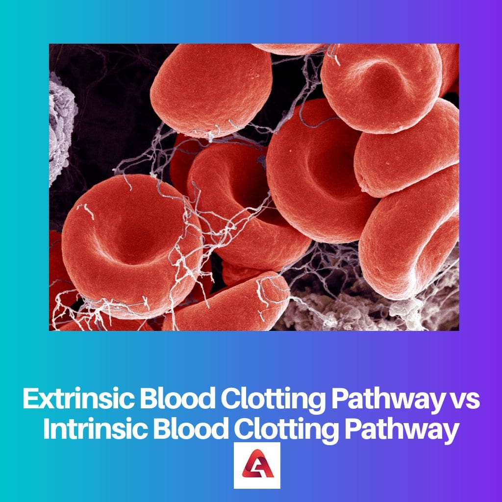
Blood clotting is a process in which the blood turns from liquid to gel form. It is a rescue step from the body to stop the blood from flowing in case of any injured veins. This leads to hemostasis. The intrinsic and extrinsic pathways activate the fibrin mesh and stabilize the platelet plug.
Key Takeaways
- The extrinsic blood clotting pathway is triggered by external factors, such as tissue damage, while the intrinsic pathway is initiated by internal factors within the bloodstream.
- The extrinsic pathway involves the activation of factor VII, while the intrinsic pathway requires the activation of factors VIII, IX, XI, and XII.
- Both extrinsic and intrinsic pathways converge at the common pathway, forming a stable blood clot.

Extrinsic Blood Clotting Pathway vs Intrinsic Blood Clotting Pathway
Any blood vessel damaged by external injury triggers the extrinsic pathway that causes blood clotting. In it, tissue factor creates clots to stop bleeding. When the glass, etc. damage the body, the intrinsic pathway helps keep blood clotting. This process involves many steps, so it is slow.
The extrinsic pathway is shorter in length. As soon as the secondary hemostasis begins, the endothelial cells release tissue factors and factor VII is activated, which then activates factor VIIa, this goes on to activate factor Xa from factor X. Clinically, the extrinsic pathway is measured to be the prothrombin time (PT).
The intrinsic pathway is the longer pathway of secondary hemostasis. The process is a cascade of activation factors. Beginning with the activation of factor XII to XIIa, this moves towards the activation of XIa factor from XI. Then factor IX activates IXa, and factor X is converted into factor Xa. In the end, all the factors are activated, and the patient positively starts blood clotting. It is measured as the partial thromboplastin time (PTT).
Comparison Table
| Parameters of Comparison | Extrinsic Blood Clotting Pathway | Intrinsic Blood Clotting Pathway |
|---|---|---|
| Length | It is shorter in length | It is longer in length |
| Factors | It consists of factors I, II, VI, and X | It consists of factors I, II, IX, X, XI, and XII |
| Triggering Point | Triggered by external trauma | Triggered by internal damage |
| Means of Activation | Activated by blood factors | Activated by tissue factors |
| Process Speed | Takes approximately 15-20 seconds to initiate blood clotting | Takes approximately 2-6 minutes to initiate blood clotting |
What is an Extrinsic Blood Clotting Pathway?
In case of an external injury that causes blood clotting is called the extrinsic blood clotting pathway. Mainly the blood factors, for example, protein, starts the process of hemostasis. The extrinsic pathway is part of secondary hemostasis.
The extrinsic pathways can be tested in the laboratory. The name of the test is Prothrombin Time. The animals which have high tissue factors are utilized to collect tissue thromboplastin.
Phospholipid, calcium, and thromboplastin are simultaneously added to the plasma that is anticoagulated with citrate buffer. The time taken to form the blood clot is called the Thromboplastin Time. It ranges from 10 to 12 seconds.
This time range during the test is compared to the normal blood clotting time. In case of any delay in the blood clotting time, the reason may be a deficiency in blood clotting factors or any chemical inhibitor that is disturbing the normal clotting time.
The two main protein cofactors, factor V and factor VIII, regulate in the blood as inactive cofactors. At the time of any injury, these cofactors become active and bind to the membrane surfaces, forming protein complexes.
Six proteins involved in blood coagulation require Vitamin K, protein C, and protein S.
What is an Intrinsic Blood Clotting Pathway?
The intrinsic pathway is initiated when an internal injury occurs, and factor XII activation follows. The factors are converted into enzymes form, and a new fibrin clot is formed.
Other than the protein factors, some negatively charged surfaces can also activate the enzyme factors. These surfaces include glass, kaolin, synthetic plastics, and fabrics.
While the inactive surfaces are oils, waxes, resins, silicones, and endothelial cells, endothelial cells are known to be the most inactive surfaces of all.
The intrinsic pathway can be tested in a laboratory through a test known as partial thromboplastin time. Plasma mixed with citrate buffer makes it anticoagulated, thus a fibrin clot can not be generated. Then a negatively charged material is added to the plasma. Resulting in the formation of membrane and, finally the clot.
The time taken during the formation of a visible blot clot is known as the partial thromboplastin time. The time ranges from 25 to 50 seconds and is then compared to the normal blood clotting time.
The intrinsic pathway is known as the longer pathway of the blood clotting process. It involves factors XII, XI, IX, and VIII. These factors are activated by platelets, exposed endothelium, or collagens.
Main Differences Between Extrinsic and Intrinsic Blood Clotting Pathways
- Extrinsic blood clotting occurs in case of external trauma, while intrinsic blood clotting occurs when there is an internal injury.
- The intrinsic pathway requires ionized calcium to activate factor IX by factor IXa, while extrinsic requires both calcium and tissue factors to activate factor IX by factor VIIa.
- The extrinsic pathway is shorter in length as compared to the intrinsic pathway.
- Blood factors activate the intrinsic pathway, while tissue factors activate the extrinsic.
- The disorder of the extrinsic pathway is referred to as Factor VII Deficiency while the disorder of the intrinsic pathway is known as Hemophilia A, B, and C. Deficiency of Factor VIII, IX, and XI, respectively.
- https://www.ncbi.nlm.nih.gov/pmc/articles/PMC5767294/
- https://journals.lww.com/bloodcoagulation/fulltext/2007/10000/Mathematical_modeling_of_blood_coagulation.9.aspx
